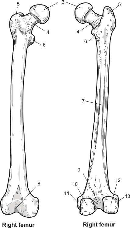
41 labelled femur
Femur bone anatomy: Proximal, distal and shaft | Kenhub The femur bone is the strongest and longest bone in the body, occupying the space of the lower limb, between the hip and knee joints. Femur anatomy is so unique that it makes the bone suitable for supporting the numerous muscular and ligamentous attachments within this region, in addition to maximally extending the limb during ambulation. femur bone anatomy not labeled knee pain tibia fibula head lateral muscle labeled schlatter osgood disease femur laser hamstrings patellar joint patella femoris chiropractor anatomy. 19 New Parts Of Femur Bone rycaaschiffman.blogspot.com. Click On A Region In The Picture To Color It In With The Selected Color.
Femur Bone - Posterior Markings - GetBodySmart Fovea of the femur head (Fovea capitis femoris) is a small, pit-like depression on the medial surface of the head.It is an attachment site for the ligamentum teres (round ligament or ligament of head of the femur). This short, narrow, ligamentous band transmits arteries to the head of the femur and helps attach the head to the acetabulum of the os coxae (hip bone).

Labelled femur
Femur Bone - Anterior Markings - GetBodySmart Solidify your knowledge of femur anatomy with these interactive quizzes and labelling exercises. 1 2 Greater trochanter ( Trochanter major) is a large, irregular-shaped process located lateral to neck and superior to shaft. It is an attachment point for the gluteus medius, gluteus minimus, piriformis, obturator, and gemellus muscles. 1 2 Labeled Femur Bone The femur bone is the strongest and longest bone in the body occupying the space of the lower limb between the hip and knee joints. This is an online quiz called Label the Femur There is a printable worksheet available for download here so you can take the quiz with pen and paper. The femurthe only bone in the upper legis a long bone. Labeled Skeletal System Diagram - Bodytomy Bone Structure of the Chest and Hip. The bones shown in the chest and hip region in the labeled human skeleton diagram are the ribs, vertebrae, pelvis, OS coxae, sacrum and coccyx. Total there are 12 pairs of ribs, as you can see in the diagram. The last pair of the ribs, which is at the bottom of the rib, are called floating ribs, as they are ...
Labelled femur. Femur Bone Anatomy: Skeletal System Lower Limb [Labeled Diagram] Anatomy of the femur, or thigh bone, made easy using a colored labeled diagram and drawing. Skeletal system anatomy of lower limb for nursing, medical learne... Femur Bone: Definition, Diagrams, Location, Parts & Functions The popliteal surface of the femur is a triangular space found at the distal posterior surface of the femur. It is bordered medially and laterally by the corresponding supracondylar lines, and inferiorly by the superior border of the fibrous capsule of the knee. The caudal aspect of the surface forms part of the floor of the popliteal fossa. Learn femur anatomy fast with these femur quizzes | Kenhub We'll begin with an overview of femur anatomy. Next, we'll look at some diagrams of the femur labeled and unlabeled to test your identification skills. Finally, we'll end with some interactive femur quizzes to fully consolidate your knowledge. Ready? Let's get into it. Unlabeled diagram showing the femur (download free PDF below!) Contents The Femur - Proximal - Distal - Shaft - TeachMeAnatomy The femur is the only bone in the thigh and the longest bone in the body. It acts as the site of origin and attachment of many muscles and ligaments, and can be divided into three parts; proximal, shaft and distal. In this article, we shall look at the anatomy of the femur - its attachments, bony landmarks, and clinical correlations. Proximal
labeled femur Flashcards | Quizlet 18 terms Kimberly_Miller33 labeled femur STUDY PLAY Articular Cartilage Canaliculi Compact bone Diaphysis Distal diaphysis Endosteum Epiphyseal plate Haversian canal Lacuna Medullary cavity Osteocyte Osteon Periosteum Proximal epiphysis Spongy bone Red marrow is found here Volkmann's canal Yellow marrow is found here Features Quizlet Live Dog Skeleton Anatomy with Labeled Diagram The segments and bones of the pelvic limb - hip, femur, tibia -fibula, tarsal, and metatarsal Identify single and paired bones from the dog skull The bones of the dog vertebrae, sternum, and ribs I hope the dog skeleton labeled diagram might help you quickly identify all the bones. › en › libraryLeg and knee anatomy: Bones, muscles, soft tissues | Kenhub Jun 09, 2022 · The main parts of the knee joint are the femur, tibia, patella, and supporting ligaments. The condyles of the femur and of the tibia come in close proximity to form the main structure of the joint. The patella, commonly known as the ‘kneecap’, is a sesamoid bone that sits within the tendon of the quadriceps femoris. It serves a protective ... Femur Labeled Diagram | Quizlet Start studying Femur Labeled. Learn vocabulary, terms, and more with flashcards, games, and other study tools.
Femur Bone Anatomy Quiz [Labeled Diagram] - YouTube Quiz yourself on the anatomy of the femur by labeling this step-by-step diagram of the main structures of the bone. Skeletal system lower extremity review fo... femur | Definition, Function, Diagram, & Facts | Britannica femur, also called thighbone, upper bone of the leg or hind leg. The head forms a ball-and-socket joint with the hip (at the acetabulum), being held in place by a ligament ( ligamentum teres femoris) within the socket and by strong surrounding ligaments. The Femur - Human Anatomy The Femur. F IG. 243- Upper extremity of right femur viewed from behind and above. (Thigh Bone) The femur (Figs. 244, 245), the longest and strongest bone in the skeleton, is almost perfectly cylindrical in the greater part of its extent. In the erect posture it is not vertical, being separated above from its fellow by a considerable interval ... Labeled Skeletal System Diagram - Bodytomy Bone Structure of the Chest and Hip. The bones shown in the chest and hip region in the labeled human skeleton diagram are the ribs, vertebrae, pelvis, OS coxae, sacrum and coccyx. Total there are 12 pairs of ribs, as you can see in the diagram. The last pair of the ribs, which is at the bottom of the rib, are called floating ribs, as they are ...
Labeled Femur Bone The femur bone is the strongest and longest bone in the body occupying the space of the lower limb between the hip and knee joints. This is an online quiz called Label the Femur There is a printable worksheet available for download here so you can take the quiz with pen and paper. The femurthe only bone in the upper legis a long bone.
Femur Bone - Anterior Markings - GetBodySmart Solidify your knowledge of femur anatomy with these interactive quizzes and labelling exercises. 1 2 Greater trochanter ( Trochanter major) is a large, irregular-shaped process located lateral to neck and superior to shaft. It is an attachment point for the gluteus medius, gluteus minimus, piriformis, obturator, and gemellus muscles. 1 2


Post a Comment for "41 labelled femur"