41 labeled diagram of a microscope
Parts of a Microscope Labeling Activity - Storyboard That Create a poster that labels the parts of a microscope and includes descriptions of what each part does. Click "Start Assignment". Use a landscape poster layout (large or small). Search for a diagram of a microscope. Using arrows and textables label each part of the microscope and describe its function. A Study of the Microscope and its Functions With a Labeled Diagram A Study of the Microscope and its Functions With a Labeled Diagram To better understand the structure and function of a microscope, we need to take a look at the labeled microscope diagrams of the compound and electron microscope. These diagrams clearly explain the functioning of the microscopes along with their respective parts. M mooketsi
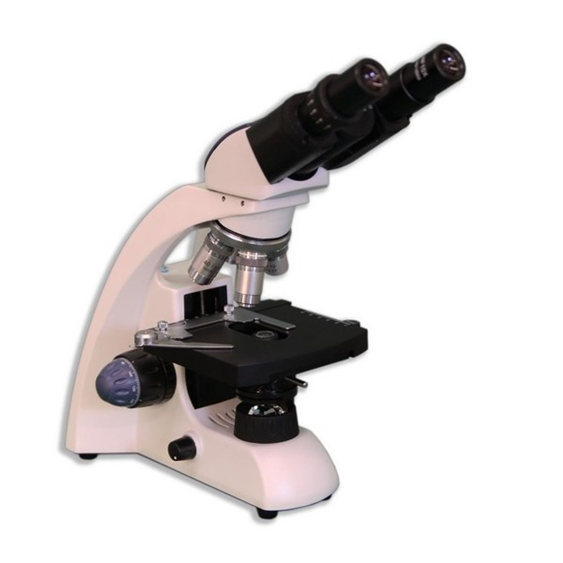
Compound Microscope Parts, Functions, and Labeled Diagram Base: Bottom base of the microscope that houses the illumination & supports the compound microscope. Objective lenses: There are usually 3-5 optical lens objectives on a compound microscope each with different magnification levels. 4x, 10x, 40x, and 100x are the most common magnifying powers used for the objectives.

Labeled diagram of a microscope
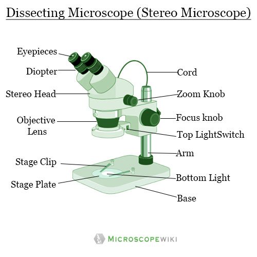
› parts-of-a-compoundMicroscope Parts and Functions With Labeled Diagram and ... Before exploring microscope parts and functions, you should probably understand that the compound light microscope is more complicated than just a microscope with more than one lens. First, the purpose of a microscope is to magnify a small object or to magnify the fine details of a larger object in order to examine minute specimens that cannot ... Cell Microscope Under Leaf Labeled Study the habits of amoebae, vorticellas, paramecium, and other protozoans under a microscope Study the habits of amoebae, vorticellas, paramecium, and other protozoans under a microscope. Label an de diagram of a stomatal apparatus 2 four parts on the diagram Find premium, high-resolution stock photography at Getty Images The students will use ... rsscience.com › stereo-microscopeParts of Stereo Microscope (Dissecting microscope) – labeled ... Labeled part diagram of a stereo microscope Major structural parts of a stereo microscope. There are three major structural parts of a stereo microscope. The viewing Head includes the upper part of the microscope, which houses the most critical optical components, including the eyepiece, objective lens, and light source of the microscope.
Labeled diagram of a microscope. Labeled Leaf Cell Microscope Under Search: Leaf Cell Under Microscope Labeled. After hundreds of years of observation, the cell theory was developed These leaves are two cells thick, so you should be able to focus up and down to see that the cells in one layer are larger than those in the other 8 Between the layers of cells inside the leaf are veins that contain xylem and After exposure to OGD for 4 h, followed by treatment ... Label the microscope — Science Learning Hub All microscopes share features in common. In this interactive, you can label the different parts of a microscope. Use this with the Microscope parts activity to help students identify and label the main parts of a microscope and then describe their functions. Drag and drop the text labels onto the microscope diagram. If you want to redo an answer, click on the box and the answer will go back to the top so you can move it to another box. Microscope Types (with labeled diagrams) and Functions Compound microscope labeled diagram. Compound microscope functions: It finds great application in areas of pathology, pedology, forensics etc; Its greater order of magnification allows for deeper study of microbial organisms to Detect the cause of diseases; Study the mineral composition in soils; Examine evidences collected in crime scenes by forensics. Compound Microscope Parts - Labeled Diagram and their Functions - Rs ... What is a "compound microscope"? Labeled diagram of a compound microscope; Major structural parts of a compound microscope; Optical components of a compound microscope. Eyepiece; Eyepiece tube; Objective lenses; Nosepiece; Specimen stage; Coarse and fine focus knobs; Rack stop; Illuminator; Condenser; Abbe condenser; Iris Diaphragm; Condenser Focus Knob; Summary
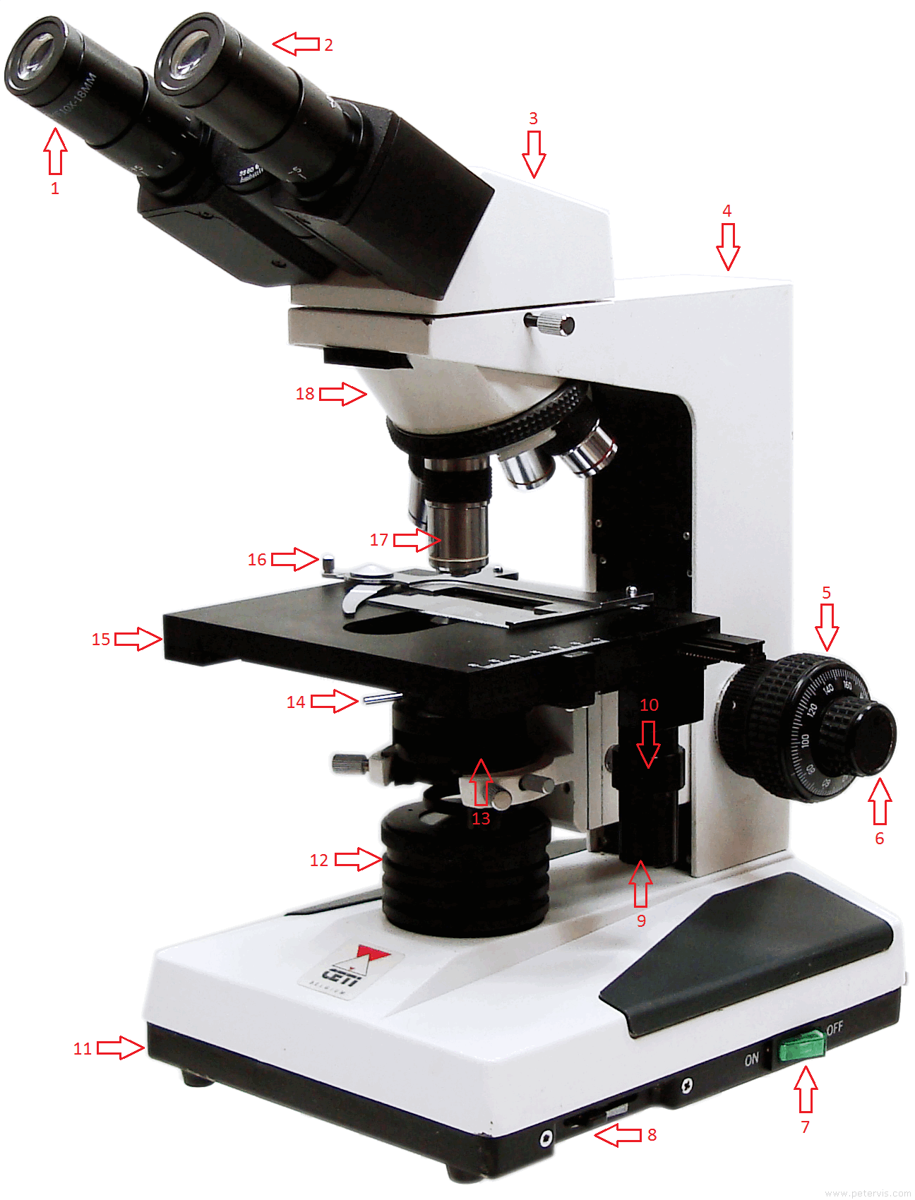
Labeling the Parts of the Microscope | Microscope World Resources Labeling the Parts of the Microscope This activity has been designed for use in homes and schools. Each microscope layout (both blank and the version with answers) are available as PDF downloads. You can view a more in-depth review of each part of the microscope here. Download the Label the Parts of the Microscope PDF printable version here. Labelled Diagram of Compound Microscope - Biology Discussion The below mentioned article provides a labelled diagram of compound microscope. Part # 1. The Stand: The stand is made up of a heavy foot which carries a curved inclinable limb or arm bearing the body tube. The foot is generally horse shoe-shaped structure (Fig. 2) which rests on table top or any other surface on which the microscope in kept. Microscope labeled diagram - SlideShare Microscope labeled diagram Oct. 30, 2013 • 6 likes • 27,530 views Download Now Download to read offline Pisgah High School Follow 1. The Microscope Image courtesy of: Microscopehelp.com Basic rules to using the microscope 1. You should always carry a microscope with two hands, one on the arm and the other under the base. 2. microbenotes.com › inverted-microscopeInverted Microscope- Definition, Principle, Parts, Labeled ... Apr 10, 2022 · The working principle of the inverted microscope is basically the same as that of an upright light microscope. They use light rays to focus on a specimen, to form an image that can be viewed by the objective lenses. However, in the inverted microscope, the light source and the condenser are found on top of the stage pointing down to the stage.
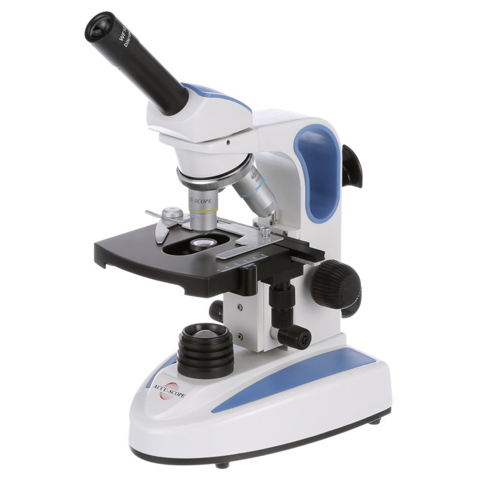
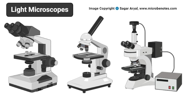
Parts of the Microscope with Labeling (also Free Printouts) Parts of the Microscope with Labeling (also Free Printouts) A microscope is one of the invaluable tools in the laboratory setting. It is used to observe things that cannot be seen by the naked eye. Table of Contents 1. Eyepiece 2. Body tube/Head 3. Turret/Nose piece 4. Objective lenses 5. Knobs (fine and coarse) 6. Stage and stage clips 7. Aperture researchtweet.com › microscope-parts-labeledMicroscope, Microscope Parts, Labeled Diagram, and Functions Jan 19, 2022 · The liquid sample comes next. To minimise evaporation and protect the microscope lens from sample exposure, a small square of clear glass or plastic (a coverslip) is placed on top of the liquid. 1. Collect a clean microscope slide and a coverslip (a thin piece of plastic covering). Fill the centre of the microscope slide with a drop or two of ... Compound Microscope: Definition, Diagram, Parts, Uses, Working ... - BYJUS Compound microscope is a type of optical microscope that is used for obtaining a high-resolution image. There are more than two lenses in a compound microscope. Learn about the working principle, parts and uses of a compound microscope along with a labeled diagram here. Light Microscope- Definition, Principle, Types, Parts, Labeled Diagram ... Figure: Labeled Diagram of a Light Microscope. Types of light microscopes (optical microscope) With the evolved field of Microbiology, the microscopes. used to view specimens are both simple and compound light microscopes, all using lenses. The difference is simple light microscopes use a single lens for magnification while compound lenses use ...
Pin di Cells & Microscopes
22 Parts Of a Microscope With Their Function And Labeled Diagram 22 Parts Of a Microscope With Their Function And Labeled Diagram Microscope Description A microscope is a laboratory instrument used to examine objects that are too small to be seen by the naked eye. In other words, it enlarges images of small objects.

Label the Microscope Diagram | Download Scientific Diagram
Label Microscope Diagram - EnchantedLearning.com Label Microscope Diagram. Using the terms listed below, label the microscope diagram. Inventions and Inventors. arm - this attaches the eyepiece and body tube to the base. base - this supports the microscope. body tube - the tube that supports the eyepiece.

Diagram of traveling microscope setup with implant cast and ...
PDF Label parts of the Microscope Label parts of the Microscope: . Created Date: 20150715115425Z

Parts of Microscope, Function, Names & Labeled Diagram ...
Simple Squamous Epithelium under a Microscope with a Labeled Diagram ... Histological features of lung parenchyma with microscopic slide images and labeled diagrams. The lung's alveoli give the honeycomb appearance in the parenchyma and lines by flattened simple squamous epithelium. These alveoli are thin-walled and fills with air. From the lung parenchyma labeled diagram, you might identify the following ...
Compound Microscope Parts, Functions, and Labeled Diagram ...
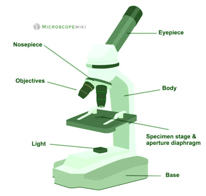
microbenotes.com › parts-of-a-microscopeParts of a microscope with functions and labeled diagram Apr 19, 2022 · Figure: Diagram of parts of a microscope. There are three structural parts of the microscope i.e. head, base, and arm. Head – This is also known as the body. It carries the optical parts in the upper part of the microscope. Base – It acts as microscopes support. It also carries microscopic illuminators.

microscope parts LABELED
A Study of the Microscope and its Functions With a Labeled Diagram A Study of the Microscope and its Functions With a Labeled Diagram To better understand the structure and function of a microscope, we need to take a look at the labeled microscope diagrams of the compound and electron microscope. These diagrams clearly explain the functioning of the microscopes along with their respective parts.

Parts of a Light Microscope - Microscopy
anatomylearner.com › cat-skeleton-anatomyCat Skeleton Anatomy with Labeled Diagram - AnatomyLearner May 29, 2021 · Cat skeleton anatomy labeled diagram. Now, I will show you all the bones from the cat skeleton with a diagram. If you find any mistakes in this cat anatomy labeled diagram, please let me know. I hope this cat skeletal system anatomy labeled diagram might help you understand and identify all the cat’s bones.

Microscope parts 3D learning for Android - APK Download
Compound Microscope - Diagram (Parts labelled), Principle and Uses See: Labeled Diagram showing differences between compound and simple microscope parts Structural Components The three structural components include 1. Head This is the upper part of the microscope that houses the optical parts 2. Arm This part connects the head with the base and provides stability to the microscope.
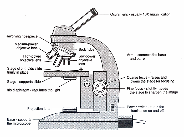
Diagram of a Microscope - Guide to using a microscope
(a) Draw the labelled ray diagram for the formation of image by a ... (a) Draw the labelled ray diagram for the formation of image by a compound microscope. Derive an expression for its total magnification (or magnifying power), when the final image is formed at the near point. (b) Why both objective and eyepiece of a compound microscope must have short focal lengths?
Microscope Types (with labeled diagrams) and Functions
thebiologynotes.com › microscopeMicroscope- Definition, Parts, Functions, Types, Diagram, Uses Feb 21, 2022 · Limitations of Dissecting Microscope or Stereo Microscope. Has limited use; Low magnification; Costly system; 8. Digital Microscope. Digital Microscope is a type of microscope that lack an ocular lens and instead contains a digital camera and screen to display image digitally. This is a modern microscope which is a computerized system combining ...

Draw a labelled diagram of a compound microscope.
Labeling Microscope Worksheet | Teaching Resources docx, 300.56 KB. A straightforward worksheet in which students are required to identify the parts of a basic microscope. Tes classic free licence.

Compound Microscope- Definition, Labeled Diagram, Principle ...
Microscope Labeling Diagram | Quizlet Unit 2 Lesson 5 - Punnett Squares and Pedigrees. 4 terms. PGFry210. Unit 2 Lesson 4 - Heredity. 9 terms. PGFry210. Upgrade to remove ads. Only $2.99/month.

Solved tration Questions: (10 points) Label the diagram of a ...
Draw a labelled diagram of an image formed by a compound microscope ... Click here👆to get an answer to your question ️ Draw a labelled diagram of an image formed by a compound microscope, with the image at least distance of distinct vision. Write any one expression for its magnifying power. ... Draw a labelled ray diagram of an image formed by a compound microscope, when the final image lies at the least ...

Compound Microscope Parts – Labeled Diagram and their ...
Parts of Microscope, Function, Names & Labeled Diagram - slidingmotion Microscope parts labeled diagram gives us all the information about its parts and their position in the microscope. Microscope Parts Labeled Diagram The principle of the Microscope gives you an exact reason to use it. It works on the 3 principles. Magnification Resolving Power Numerical Aperture. Parts of Microscope Head Base Arm Eyepiece Lens
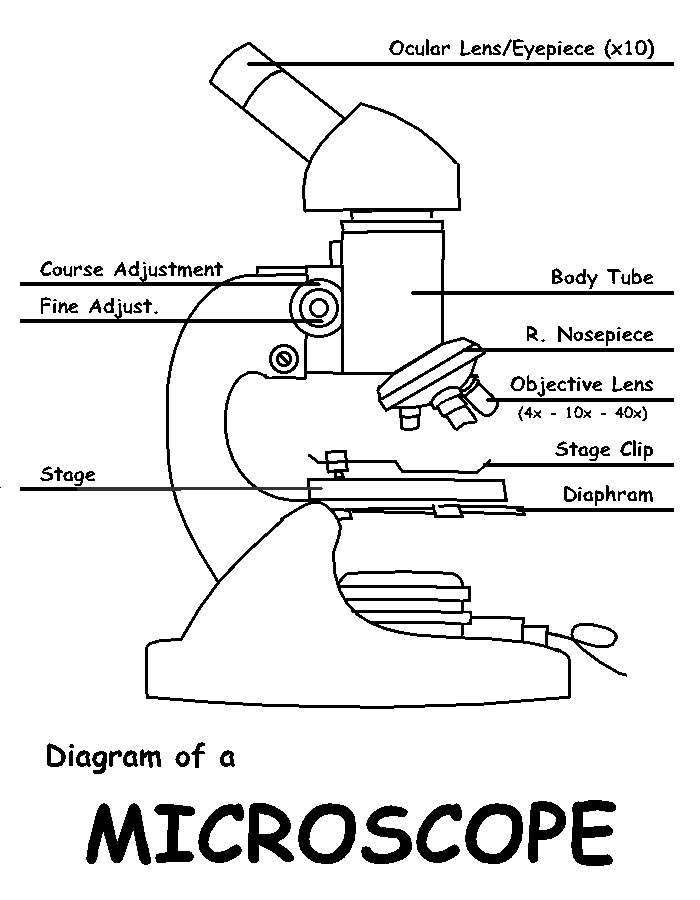
Diagram of a Microscope by ScienceDoodles on DeviantArt
rsscience.com › stereo-microscopeParts of Stereo Microscope (Dissecting microscope) – labeled ... Labeled part diagram of a stereo microscope Major structural parts of a stereo microscope. There are three major structural parts of a stereo microscope. The viewing Head includes the upper part of the microscope, which houses the most critical optical components, including the eyepiece, objective lens, and light source of the microscope.
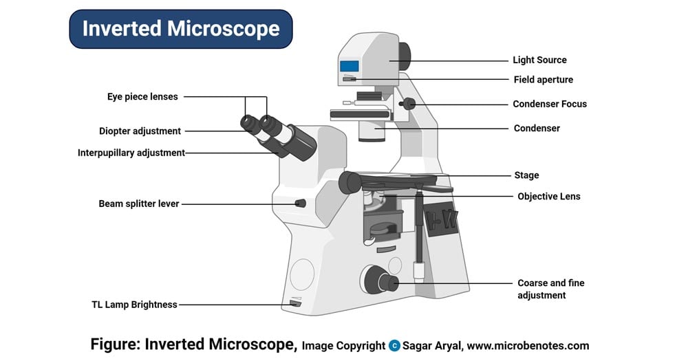
Inverted Microscope- Definition, Principle, Parts, Labeled ...
Cell Microscope Under Leaf Labeled Study the habits of amoebae, vorticellas, paramecium, and other protozoans under a microscope Study the habits of amoebae, vorticellas, paramecium, and other protozoans under a microscope. Label an de diagram of a stomatal apparatus 2 four parts on the diagram Find premium, high-resolution stock photography at Getty Images The students will use ...

Compound Microscope Parts – Labeled Diagram and their ...
› parts-of-a-compoundMicroscope Parts and Functions With Labeled Diagram and ... Before exploring microscope parts and functions, you should probably understand that the compound light microscope is more complicated than just a microscope with more than one lens. First, the purpose of a microscope is to magnify a small object or to magnify the fine details of a larger object in order to examine minute specimens that cannot ...

Microscope With Labels Clip Art at Clker.com - vector clip ...

Parts of a Microscope Labeling Activity
Dissecting microscope (Stereoscopic or stereo microscope ...

Compound Microscope – Diagram (Parts labelled), Principle and ...
Labelled Diagram of Microscope Parts
Compound Microscope Parts, Functions, and Labeled Diagram ...

Microscopes: A Beginner's Guide

Labeling the Parts of the Microscope | Microscope World Resources

Diagram of Compound Microscope
Light Microscope- Definition, Principle, Types, Parts ...
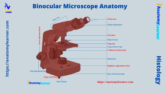
Binocular Microscope Anatomy - Parts and Functions with a ...

Labeled Diagram of Microscope Clipart Free Download

give a well labelled diagram of compound microscope using of ...
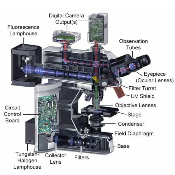
Fluorescence Microscopy - Explanation and Labelled Images ...
Junior cert Labelling Microscope - Labelled diagram

Unlabeled Microscope Diagram posted by Christopher Thompson

Parts of Stereo Microscope (Dissecting microscope) – labeled ...
Labeled-microscope-diagram

File:Microscope diagram.png - Wikimedia Commons

How to Draw a Microscope and Label Its Parts
Simple Microscope - Diagram (Parts labelled), Principle ...

File:Labelledmicroscope.gif - Wikibooks, open books for an ...

Microscope Labeling Diagram | Quizlet

Compound Microscope Parts, Functions, and Labeled Diagram ...
Post a Comment for "41 labeled diagram of a microscope"