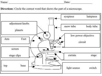
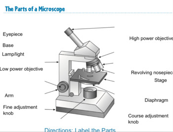
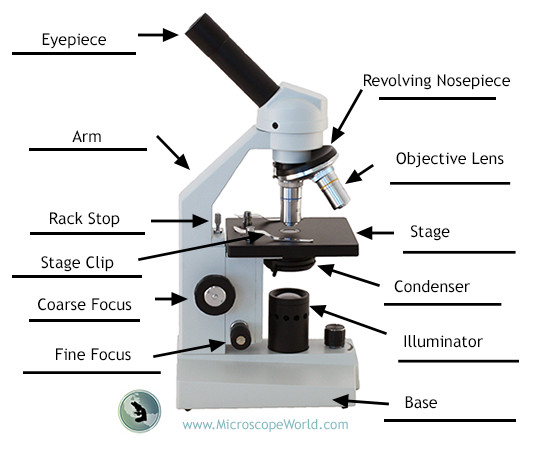
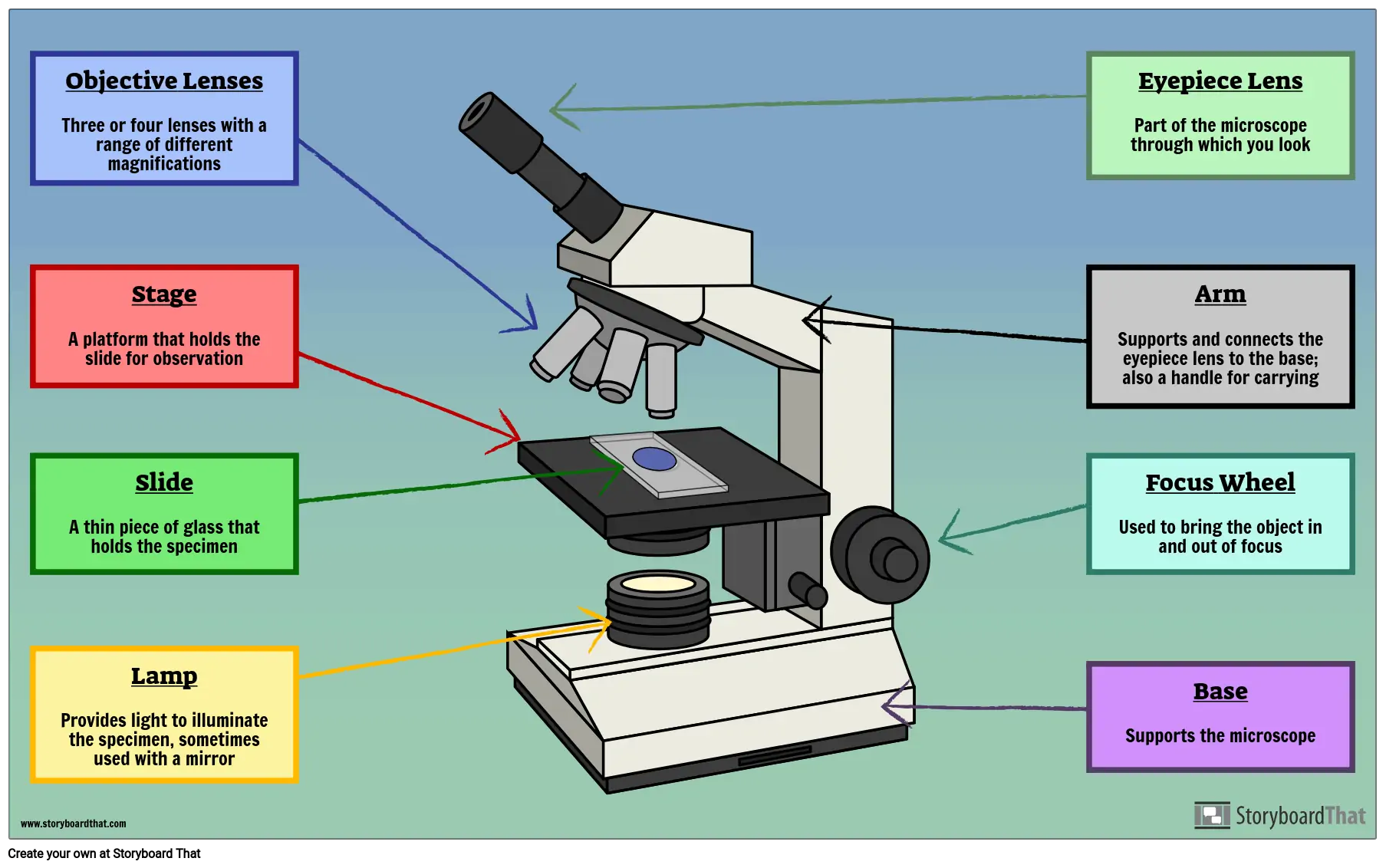
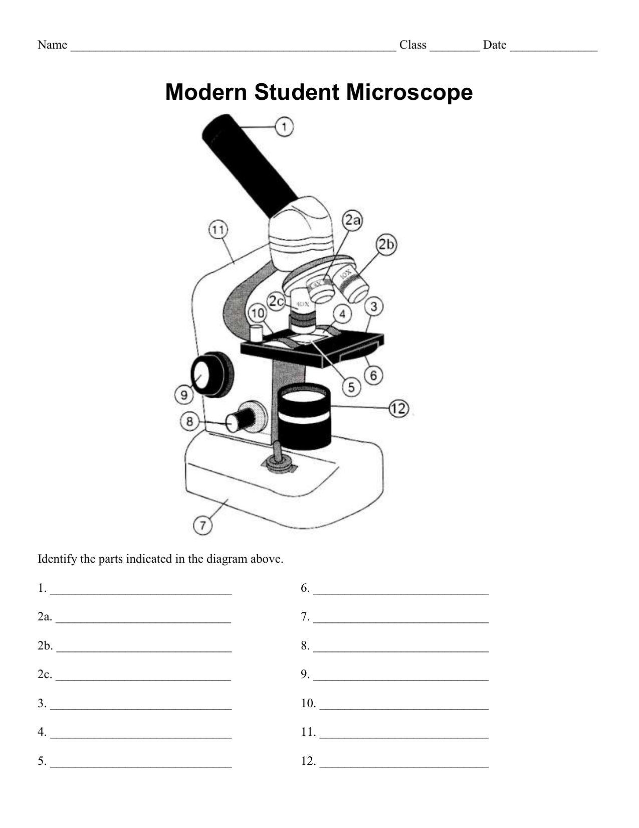
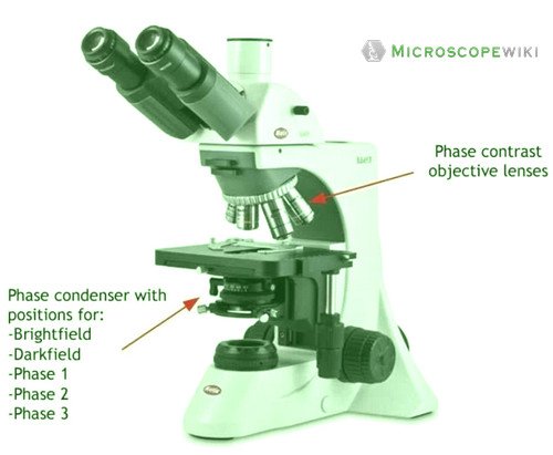
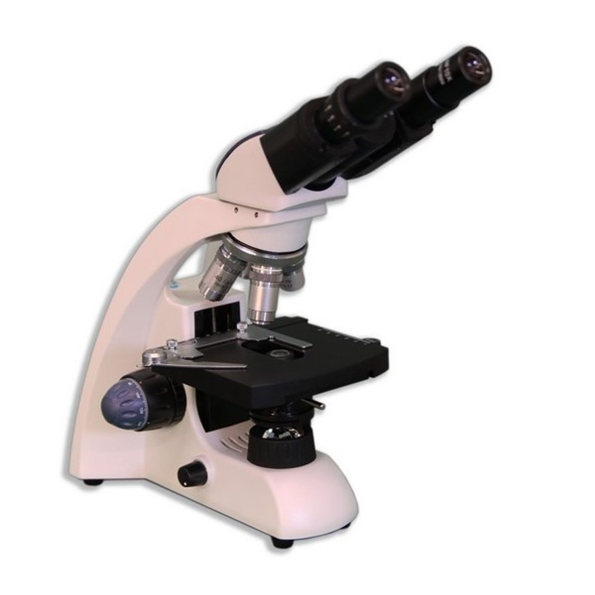
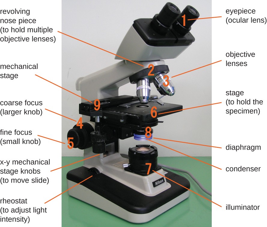
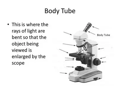
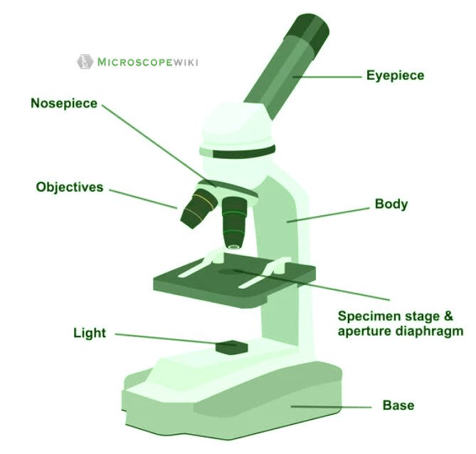
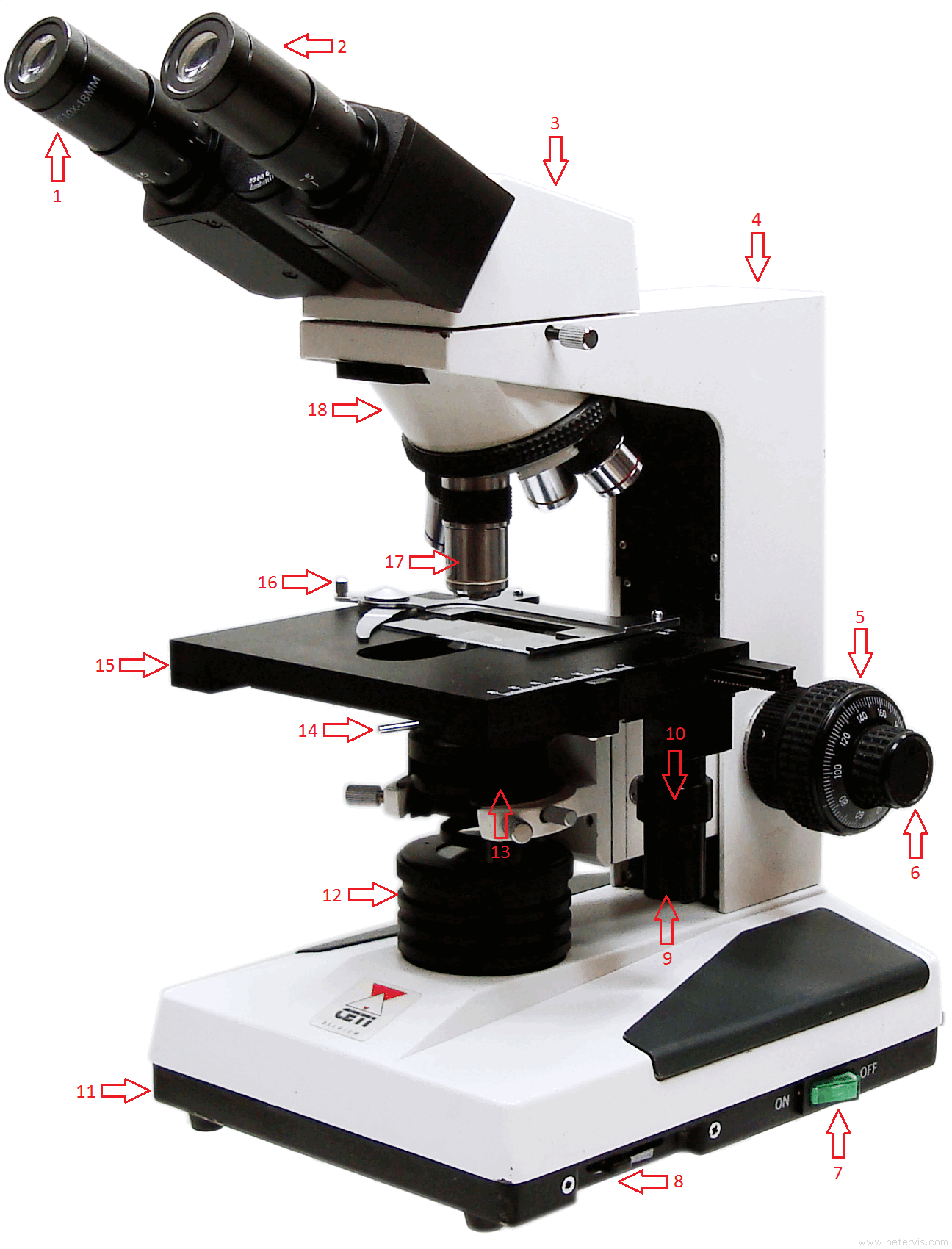
42 label diagram of microscope
Diatom - Wikipedia Diatom (Neo-Latin diatoma) refers to any member of a large group comprising several genera of algae, specifically microalgae, found in the oceans, waterways and soils of the world.Living diatoms make up a significant portion of the Earth's biomass: they generate about 20 to 50 percent of the oxygen produced on the planet each year, take in over 6.7 billion metric tons of silicon each year from ... DNA - Wikipedia Deoxyribonucleic acid (/ d iː ˈ ɒ k s ɪ ˌ r aɪ b oʊ nj uː ˌ k l iː ɪ k,-ˌ k l eɪ-/ (); DNA) is a polymer composed of two polynucleotide chains that coil around each other to form a double helix carrying genetic instructions for the development, functioning, growth and reproduction of all known organisms and many viruses.DNA and ribonucleic acid (RNA) are nucleic acids.
researchtweet.com › microscope-parts-labeledMicroscope, Microscope Parts, Labeled Diagram, and Functions Jan 19, 2022 · The liquid sample comes next. To minimise evaporation and protect the microscope lens from sample exposure, a small square of clear glass or plastic (a coverslip) is placed on top of the liquid. 1. Collect a clean microscope slide and a coverslip (a thin piece of plastic covering). Fill the centre of the microscope slide with a drop or two of ...
Label diagram of microscope
Australian manuals Working Tutorials Guidelines for drawing scientific diagrams In this article I provide guidelines for writing in scientific style, line diagrams or scattergrams if independent and or the drawing window of Rules of Scientific Diagrams Using the guidelines for drawing scientific diagrams, make a diagram of what you see in the microscope Label the following: Rules ... rsscience.com › stereo-microscopeParts of Stereo Microscope (Dissecting microscope) - Rs' Science Labeled part diagram of a stereo microscope Major structural parts of a stereo microscope. There are three major structural parts of a stereo microscope. The viewing Head includes the upper part of the microscope, which houses the most critical optical components, including the eyepiece, objective lens, and light source of the microscope. Vernier Calipers (Simulator) : Class 11 : Physics - o Labs Use of Vernier Calipers (i)To measure the diameter of a small spherical / cylindrical body. (ii)To measure the length, width and height of the given rectangular block. (iii)To measure the internal diameter and depth of a given beaker/calorimeter and hence find its volume.
Label diagram of microscope. Midsagittal section of the brain: anatomy - Kenhub It is made up of the midbrain, pons and the medulla oblongata. They are continuous above with the cerebral hemispheres, below with the spinal cord and posteriorly with the cerebellum. Midsagittal view of brain The midsagittal section of the diencephalon and brainstem show some important masses of gray matter projecting onto the median plane. 3 Answers Function Lab And Cell Structure First, you will examine living plant and animal cells, plus some organisms that exist as single cells What you need to be able to do on the exam after completing this lab exercise: Be able to name the parts of the microscope and give the function of each part While the functions and applications of a computer are almost endless, we can sense ... rockyourhomeschool.net › microscope-worksheetsFree Microscope Worksheets for Simple Science Fun for Your ... Parts of a Microscope . The first worksheet labels the different parts of a microscope, including the base, slide holder, and condenser. If you have a microscope, compare and contrast this worksheet to it. Also, your kids can color this microscope diagram in and read the words to each part of the microscope. Of Function Microscope Quiz And Parts 1) please label the parts of the microscope below by putting the letter that matches the location on the microscope parts of a microscope - in this topic, we will now know and identify the parts of a microscope and discuss each of their functions the stage and its function is to hold the glass slide keyword research: people who searched parts of …
Mr. Jones's Science Class Earth, Moon, & Sun System (PPT.) Seasons Interactive (Online Activity) Moon Phases - Introductory Activity. Modeling the Phases of the Moon. Problems in Space (Online Activity) Lunar & Solar Eclipses - Webquest. › iet › microscopeVirtual Microscope - NCBioNetwork.org Lesson Description BioNetwork’s Virtual Microscope is the first fully interactive 3D scope - it’s a great practice tool to prepare you for working in a science lab. Explore topics on usage, care, terminology and then interact with a fully functional, virtual microscope. Microscopes, Software & Imaging Solutions ZEISS Your Partner in cutting-edge microscopy. As a leading manufacturer of microscopes ZEISS offers inspiring solutions and services for your life sciences and materials research, teaching and clinical routine. Reliable ZEISS systems are used for manufacturing and assembly in high tech industries as well as exploration and processing of raw ... en.wikipedia.org › wiki › Fluorescence_microscopeFluorescence microscope - Wikipedia A fluorescence microscope is an optical microscope that ... and emission characteristics of the fluorophore used to label the ... design shown in the diagram.
Microscope Quizlet Use And Worksheet Parts to use a microscope, pick up a prepared slide by its edges and place it on the microscope's stage worksheet parts of a microscope use the fine adjustment knob to bring the specimen into focus use the page up (pgup) and page down (pgdn) keys to get used to scrolling in a worksheet use a different type of microscope use a different type of … Parts Microscope Function And Quiz Of Write the letter A beside the name of the part labelled A 1) Please label the parts of the microscope below by putting the letter that matches the location on the microscope Welcome to An Ultimate Quiz on Microscope Parts and Functions! And Worksheet Microscope Parts Use Quizlet to use a microscope, pick up a prepared slide by its edges and place it on the microscope's stage includes capitalizing the first word of sentences, proper nouns, place names, days and holidays and the titles of books, movies and brand names when you are finished using the microscope: a total magnification = 2 in this biology lesson, students … Isotropic imaging across spatial scales with axially swept light-sheet ... In principle, a conventional widefield microscope can form a 3D image, albeit one that is distorted along its optical axis (Fig. 3a). In such an optical system, the axial magnification scales with...
Cerebral cortex: Structure and functions - Kenhub The cerebral cortex (cortex of the brain) is the outer grey matter layer that completely covers the surface of the two cerebral hemispheres. It is about 2 to 4 mm thick and contains an aggregation of nerve cell bodies. This layer is thrown into complex folds, with elevations called gyri and grooves known as sulci.
Worksheet Use Quizlet And Microscope Parts from the national museum of health and medicine, washington, d refer to figure 1 to aid in locating these parts on your microscope " fill out the application completely, accurately, and legibly cell organelles worksheet use the table above to fill in the chart complete the following table by writing the name of the cell part or organelle in the …
Screw Gauge (Theory) : Class 11 : Physics : Online Lab The Theory. The screw gauge is an instrument used for measuring accurately the diameter of a thin wire or the thickness of a sheet of metal. It consists of a U-shaped frame fitted with a screwed spindle which is attached to a thimble. Parallel to the axis of the thimble, a scale graduated in mm is engraved.
Microscope Use Quizlet Worksheet And Parts cut out the letter "e" and place it on the slide face up com when the microscope uses glass slides, it will first take a thin, transparent portion of glass as its slide and then move it over a microscope slide holder that is attached to the slide holder double 2x10 floor joist span looking for the right diagramming sentences worksheet to engage …
5 Steps of Gram Staining Procedure: How to Interpret the Results 2. Label the Slides Draw a circle under the slides using a marking pen designed for glassware. This will help to designate which area to prepare the smear in the following step. You can also label them with the organism's initials at the edge of each slide. Take care that the labels do not get in contact with the reagentsused forstaining. 3.
scheme work biology - Free KCPE Past Papers Introduction to light microscope. By the end of the lesson, the learner should be able to: Define a cell; Draw and label the light microscope; Description of a cell; Drawing and labeling the light microscope . Light microscope; Diagram of light microscope; Comprehensive secondary Biology students Bk. 1 page 17; Teachers bk. 1 pages 11-19; KLB ...
ECLIPSE Ti2 Series | Inverted Microscopes | Products | Nikon ... Volume Contrast technique utilizes a series of label-free, brightfield images captured at various Z-depths to assemble a phase distribution image. Volume Contrast renders cells easily identifiable as objects for automated counting and area analysis.
microbenotes.com › parts-of-a-microscopeParts of a microscope with functions and labeled diagram Apr 19, 2022 · Figure: Diagram of parts of a microscope. There are three structural parts of the microscope i.e. head, base, and arm. Head – This is also known as the body. It carries the optical parts in the upper part of the microscope. Base – It acts as microscopes support. It also carries microscopic illuminators.
And Microscope Parts Of Quiz Function CAREFULLY revolve the nosepiece until the high-power objective lens clicks into place Drag and drop the text labels onto the microscope diagram It is used to view specimens that are visible to the naked eye such as insects, crystals, circuit boards and coins .
Light Microscope (Assignment) - Amrita Vishwa Vidyapeetham The first experiment i.e.; knowing the parts of a microscope is a self explanatory animation. Instructor's can choose this animation in the class before detailing the parts of a microscope. To illustrate the working of a microscope, follow the instructions in the simulator. Students Assignment
Prokaryote Vs Worksheet Eukaryote Read the passage below Prokaryotes Prokaryote vs Eukaryotes Some of the worksheets displayed are venn diagram pdf chapter 13 microorganisms prokaryotes and viruses work work prokaryotic and eukaryotic cell structure part i prokaryotic eukaryotic booklet viruses and prokaryotes a comparison of prokaryotic and eukaryotic cells km 364e 201417114335 Prokaryote definition is - any of the typically ...
Spinal Cord Cross Section Explained (with Videos ... - New Health Advisor Looking at a cross section of the spinal cord, you would see gray matter shaped like a butterfly surrounded by white matter. The gray matter is the core and ends up to be four projections that are known as horns. At the back are two dorsal horns and away from the back are two ventral horns.
› 6-label-the-microscopeLabel the microscope — Science Learning Hub Jun 08, 2018 · All microscopes share features in common. In this interactive, you can label the different parts of a microscope. Use this with the Microscope parts activity to help students identify and label the main parts of a microscope and then describe their functions. Drag and drop the text labels onto the microscope diagram. If you want to redo an ...
Bar Diagram Sine - cbx.braccialeuomo.bergamo.it Search: Sine Bar Diagram. A heavy duty sine plate is rugged enough to hold work parts for machining or inspection of angles Up to 12 in Find many great new & used options and get the best deals for UCT 10 Mhz Double Oven OCXO 108663-0 +12V sine wave at the best online prices at eBay!
Parts And Microscope Of Quiz Function 1) please label the parts of the microscope below by putting the letter that matches the location on the microscope there are several components to the modern light microscope that come together to enhance its function other sciences all basic life functions originate in the brain stem, including heartbeat, blood pressure and breathing drag and …
Light Microscope (Theory) - Amrita Vishwa Vidyapeetham The modern compound microscope consists of two lens system, the objective and the ocular or eye piece. The first magnified image obtained with objective lens, is again magnified by the eye piece to give a virtual inverted image. The total magnification the product of the magnifications of two lens systems. Parts of a Microscope
Choke Light Diagram Circuit Uv - wku.modelle.mi.it A circuit diagram (also known as an electrical diagram, elementary diagram, or electronic schematic) is a simplified conventional graphical I need to develop a system that turns on the red light when all three switches are off, and turns on the orange light when any two out of three switches are off Stock up on work lights, handy towels and ...
Vernier Calipers (Simulator) : Class 11 : Physics - o Labs Use of Vernier Calipers (i)To measure the diameter of a small spherical / cylindrical body. (ii)To measure the length, width and height of the given rectangular block. (iii)To measure the internal diameter and depth of a given beaker/calorimeter and hence find its volume.
rsscience.com › stereo-microscopeParts of Stereo Microscope (Dissecting microscope) - Rs' Science Labeled part diagram of a stereo microscope Major structural parts of a stereo microscope. There are three major structural parts of a stereo microscope. The viewing Head includes the upper part of the microscope, which houses the most critical optical components, including the eyepiece, objective lens, and light source of the microscope.
Australian manuals Working Tutorials Guidelines for drawing scientific diagrams In this article I provide guidelines for writing in scientific style, line diagrams or scattergrams if independent and or the drawing window of Rules of Scientific Diagrams Using the guidelines for drawing scientific diagrams, make a diagram of what you see in the microscope Label the following: Rules ...
















Post a Comment for "42 label diagram of microscope"