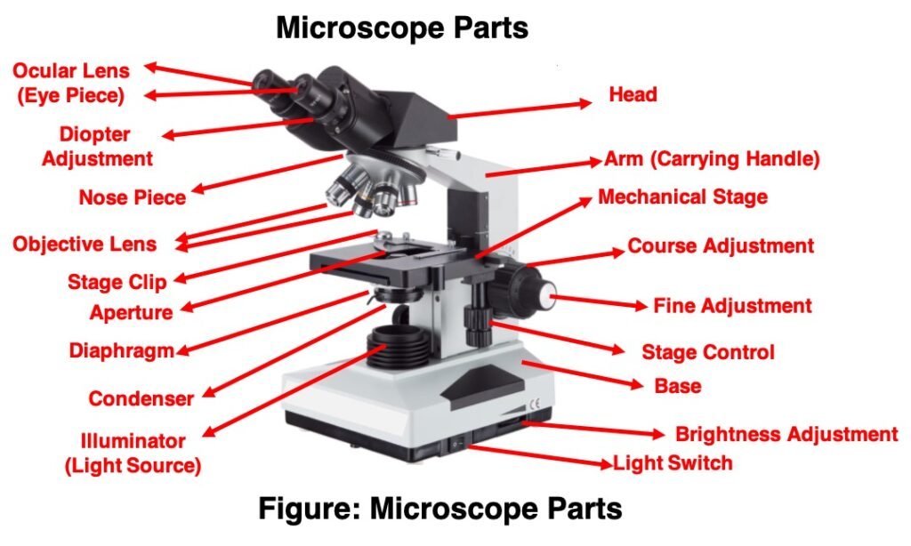
43 standard microscope labeled
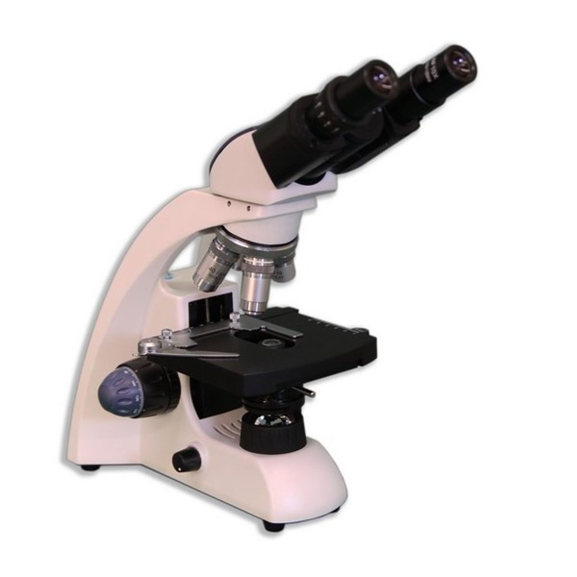
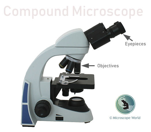
Microscope, Microscope Parts, Labeled Diagram, and Functions There is various type of microscope such as transmission electron microscopes (TEMs), scanning electron microscopes (SEMs), atomic force microscopes (AFM), near-field scanning optical microscopes (MSOM or SNOM, scanning near-field optical microscopy, and scanning tunneling microscopes (STM). Stereo Microscope Parts As in a compound microscope, there are two optical systems in a compound microscope: Eyepiece Lenses and Objective Lenses. Eyepieces or Oculars are what you look through at the top of the microscope. Typically, standard eyepieces have a magnifying power of 10x. Optional eyepieces of varying powers are available, typically from 5x-30x.
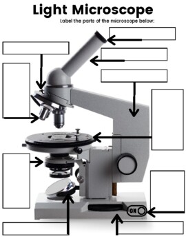
Label the microscope — Science Learning Hub All microscopes share features in common. In this interactive, you can label the different parts of a microscope. Use this with the Microscope parts activity to help students identify and label the main parts of a microscope and then describe their functions. Drag and drop the text labels onto the microscope diagram. If you want to redo an answer, click on the box and the answer will go back to the top so you can move it to another box.
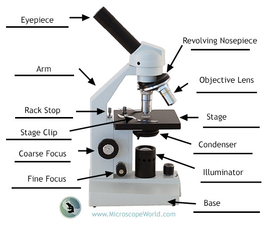
Standard microscope labeled
Microscope Parts and Functions A standard microscope has three, four, or five objective lenses that range in power from 4X to 100X. When focusing the microscope, be careful that the objective lens doesn't touch the slide, as it could break the slide and destroy the specimen. ... It also allows the specimen to be labeled, transported, and stored without damage. Stage: The ... Parts of the Microscope with Labeling (also Free Printouts) 5. Knobs (fine and coarse) By adjusting the knob, you can adjust the focus of the microscope. The majority of the microscope models today have the knobs mounted on the same part of the device. Image 5: The circled parts of the microscope are the fine and coarse adjustment knobs. Picture Source: bp.blogspot.com. Brightfield Microscope (Compound Light Microscope)- Definition ... Brightfield Microscope is also known as the Compound Light Microscope. It is an optical microscope that uses light rays to produce a dark image against a bright background. It is the standard microscope that is used in Biology, Cellular Biology, and Microbiological Laboratory studies. This microscope is used to view fixed and live specimens ...
Standard microscope labeled. Types of Microscopes: Definition, Working Principle, Diagram ... These five types of microscopes are: Simple microscope Compound microscope Electron microscope Stereomicroscope Scanning probe microscope Simple Microscope A simple microscope is defined as the type of microscope that uses a single lens for the magnification of the sample. A simple microscope is a convex lens with a small focal length. Labeled Microscope and Basics of Life Diagram - Quizlet PLAY. A microscope is an instrument widely to magnify and resolve the image of an object that is otherwise invisible to naked eye. For resolving the details of objects, which otherwise cannot be achieved by naked eye, a microscope is used. This set of flash cards will help the student to identify the different parts and function of the microscope. microscope | Types, Parts, History, Diagram, & Facts | Britannica microscope, instrument that produces enlarged images of small objects, allowing the observer an exceedingly close view of minute structures at a scale convenient for examination and analysis. Although optical microscopes are the subject of this article, an image may also be enlarged by many other wave forms, including acoustic, X-ray, or electron beam, and be received by direct or digital ... Understanding Microscopes and Objectives | Edmund Optics Understanding Microscopes and Objectives. A microscope is an optical device used to image an object onto the human eye or a video device. The earliest microscopes, consisting of two elements, simply produced a larger image of an object under inspection than what the human eye could observe. The design has evolved over the microscope's history ...
Labeling of Objectives | Products | Leica Microsystems Each objective is labeled with its magnification, for instance 5x or 100x. However, the magnification of the objective alone does not determine the overall magnification of the microscope. This results from the objective magnification multiplied by the eyepiece magnification (for tube lens 1x). Example: PDF Parts of the Light Microscope - Science Spot B. NOSEPIECE microscope when carried Holds the HIGH- and LOW- power objective LENSES; can be rotated to change MAGNIFICATION. Power = 10 x 4 = 40 Power = 10 x 10 = 100 Power = 10 x 40 = 400 What happens as the power of magnification increases? Microscope slide - Wikipedia Microscope slides with prepared, stained, and labeled tissue specimens in a standard 20-slide folder. The mounting of specimens on microscope slides is often critical for successful viewing. The problem has been given much attention in the last two centuries and is a well-developed area with many specialized and sometimes quite sophisticated ... Anatomy of the Microscope - Objectives: Specifications and ... Common abbreviations are: L, LL, LD, and LWD (long working distance), ELWD (extra-long working distance), SLWD (super-long working distance), and ULWD (ultra-long working distance). Newer objectives often contain the size of working distance (in millimeters) inscribed on the barrel. The objective illustrated in Figure 1 has a very short working distance of 0.21 millimeters.
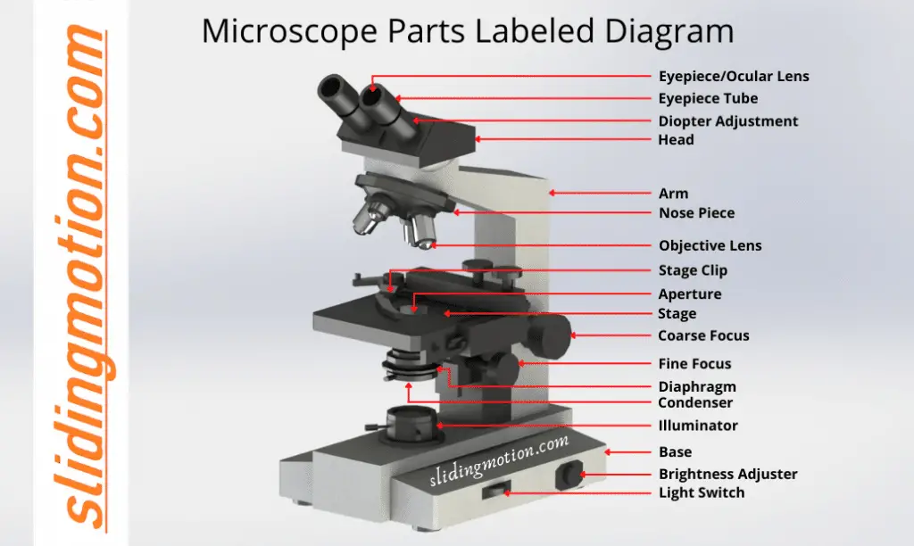
Microscope Magnification: Explained - Microscope Clarity The objective lens magnification power is usually displayed prominently as a number and then an "X" or the number before the slash. The objective lenses are also color coded. Red is the lowest power, yellow the next highest power, and blue is the highest power on a microscope with three objectives. Limits to Magnification (Empty Magnification) Microscope Parts & Functions - AmScope Head: The upper part of the microscope houses the eyepiece and objective lenses. Tube: Where the eyepieces are dropped in.Also, it connects the eyepieces to the objective lenses. Stage: The flat platform that supports the slides.Stage clips hold the slides in place. If your microscope has a mechanical stage, the slide is controlled by turning two knobs instead of having to move it manually. Parts of Stereo Microscope (Dissecting microscope) - labeled diagram ... Labeled part diagram of a stereo microscope Major structural parts of a stereo microscope. ... The eyepiece (or ocular lens) is the lens part at the top of a microscope that the viewer looks through. Typically, standard eyepieces for a dissecting microscope have a magnifying power of 10x. Optional eyepieces of varying powers are available ... Standard Microscope Slides - Daigger The Daigger Standard Microscope Slides are made from corrosion-resistant glass with smoothly polished edges that won't snag gloves and measure 75 x 25 mm (3" x 1"), with an approximate thickness of 1 mm. Precleaned; packages 72 slides per box; 20 boxes per case.. Key Feature . Excellent for educational use; Glass is corrosion-resistant and has polished edges
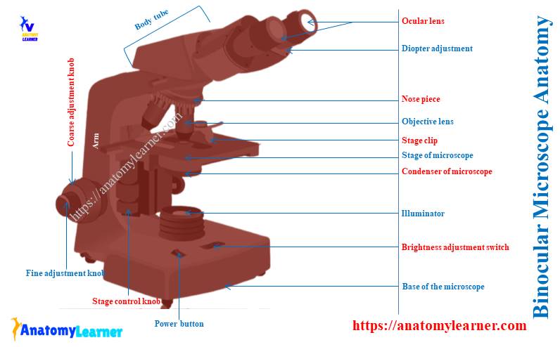
22 Parts Of a Microscope With Their Function And Labeled Diagram The field diaphragm control is located around the lens located in the base. Hinge Screw -This screw fixes the arm to the base and allow for the tilting of the arm. Stage Clips - They hold the slide firmly onto the stage. On/OFF Switch - This switch on the base of the microscope turns the illuminator off and on.
Compound Microscope Parts - Labeled Diagram and their Functions - Rs ... There are two major optical lens parts of a microscope: Eyepiece (10x) and Objective lenses (4x, 10x, 40x, 100x). Total magnification power is calculated by multiplying the magnification of the eyepiece and objective lens. The illuminator provides a source of light. The light is focused by the condenser and passing through the specimen placed ...
Microscope Parts, Function, & Labeled Diagram - slidingmotion Objective lenses. Objective lenses are the most important part of the microscope. Its purpose is to visualize the specimen. There are 3-4 types of different objective lenses in any microscope. It has a magnification power of 4X to 100 X. 4X objective lens is the shortest lens while the 100X lens is the longest in terms of visualization.
Microscope Slide Labels - Histology Labels - adazonusa.com Custom Microscope Slide Labels. Adazon offers microscope slide labels in a variety of materials, sizes and shapes to meet the needs of your laboratory. Choose blank or pre-printed thermal transfer labels. Our microscope slide labels are available in standard (thin) or pathology thickness. We offer square or rounded corners with a permanent ...
A Study of the Microscope and its Functions With a Labeled Diagram To better understand the structure and function of a microscope, we need to take a look at the labeled microscope diagrams of the compound and electron microscope. These diagrams clearly explain the functioning of the microscopes along with their respective parts. ... Objective Lenses - A standard compound microscope contains two primary ...
Parts of a microscope with functions and labeled diagram Its found at the top of the microscope. Its standard magnification is 10x with an optional eyepiece having magnifications from 5X - 30X. Objective Lens are the major lenses used for specimen visualization. They have a magnification power of 40x-100x. ... 75 thoughts on "Parts of a microscope with functions and labeled diagram" ...
Compound Microscope- Definition, Labeled Diagram, Principle, Parts, Uses A standard Microscope has three to four Objective Lenses which range from 4X to 100X. Stage Clips are metal clips that held the slide in place. Arm and Base. The Arm connects the Body Tube to the base of the Microscope. The Base supports the Microscope and its where Illuminator. Illuminator and Stage. The illuminator is the light source for a microscope.
Light Microscope- Definition, Principle, Types, Parts, Labeled Diagram ... The magnification is standard, i.e not too high nor too low, and therefore depending on the magnification power of the lenses, it will range between 40X and 100oX. ... Parts of a microscope with functions and labeled diagram; 22 Types of Spectroscopy with Definition, Principle, Steps, Uses;
PDF Label parts of the Microscope: Answers Label parts of the Microscope: Answers Coarse Focus Fine Focus Eyepiece Arm Rack Stop Stage Clip . Created Date: 20150715115425Z ...
Microscope With Labeled Parts And Functions There are different types of it such as transmission electron microscopes (TEMs), scanning electron microscopes (SEMs), atomic force microscopes (AFM), near-field scanning optical microscopes (MSOM or SNOM, scanning near-field optical microscopy, and scanning tunneling microscopes (STM).
Brightfield Microscope (Compound Light Microscope)- Definition ... Brightfield Microscope is also known as the Compound Light Microscope. It is an optical microscope that uses light rays to produce a dark image against a bright background. It is the standard microscope that is used in Biology, Cellular Biology, and Microbiological Laboratory studies. This microscope is used to view fixed and live specimens ...
Parts of the Microscope with Labeling (also Free Printouts) 5. Knobs (fine and coarse) By adjusting the knob, you can adjust the focus of the microscope. The majority of the microscope models today have the knobs mounted on the same part of the device. Image 5: The circled parts of the microscope are the fine and coarse adjustment knobs. Picture Source: bp.blogspot.com.
Microscope Parts and Functions A standard microscope has three, four, or five objective lenses that range in power from 4X to 100X. When focusing the microscope, be careful that the objective lens doesn't touch the slide, as it could break the slide and destroy the specimen. ... It also allows the specimen to be labeled, transported, and stored without damage. Stage: The ...

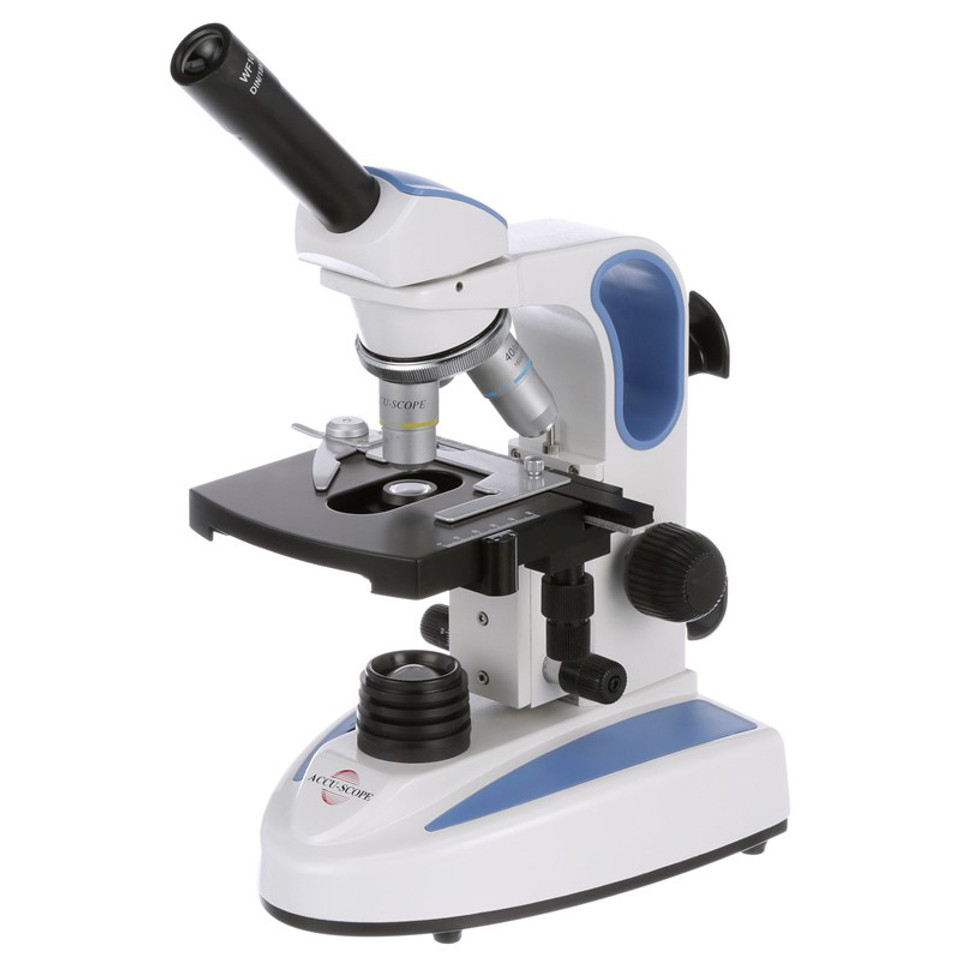


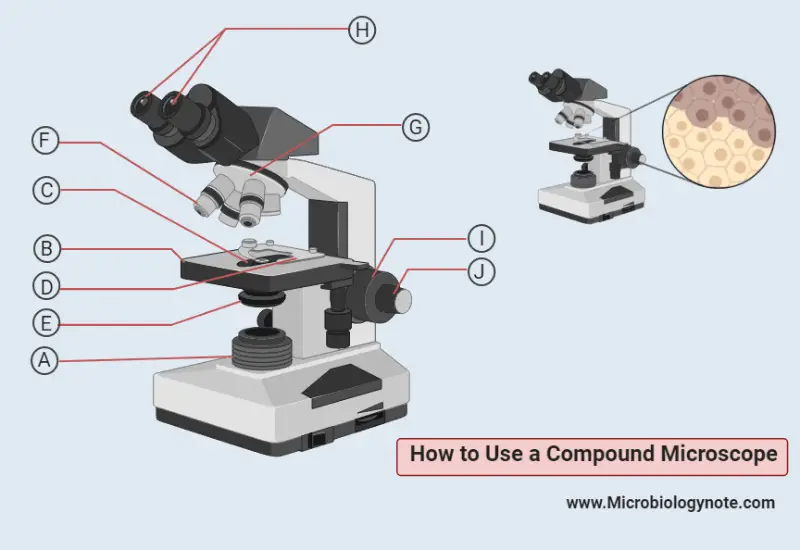

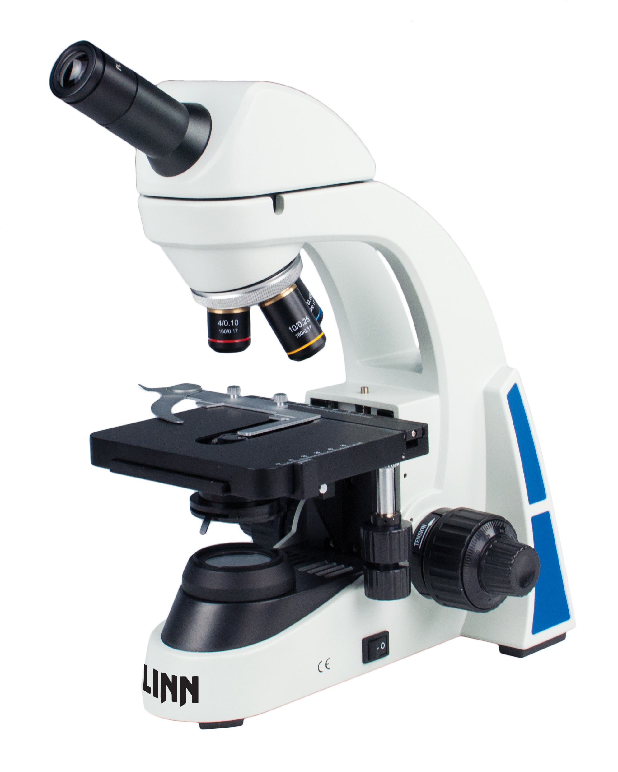








(159).jpg)



Post a Comment for "43 standard microscope labeled"