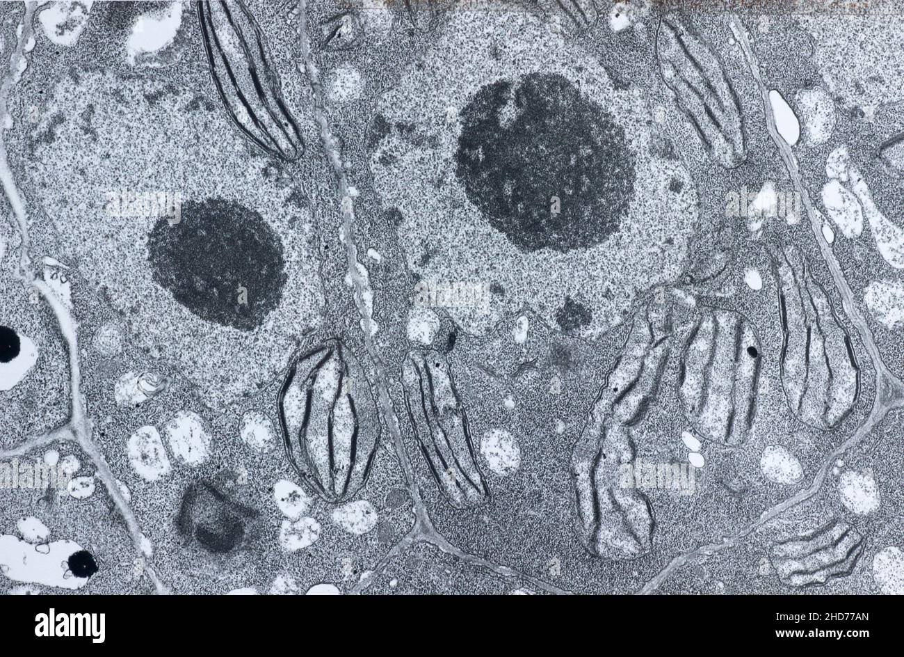
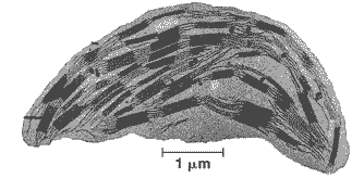
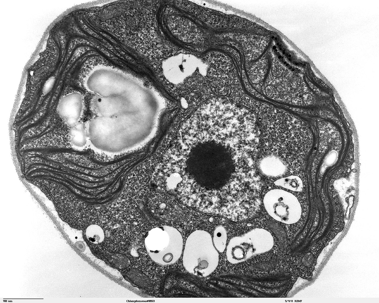
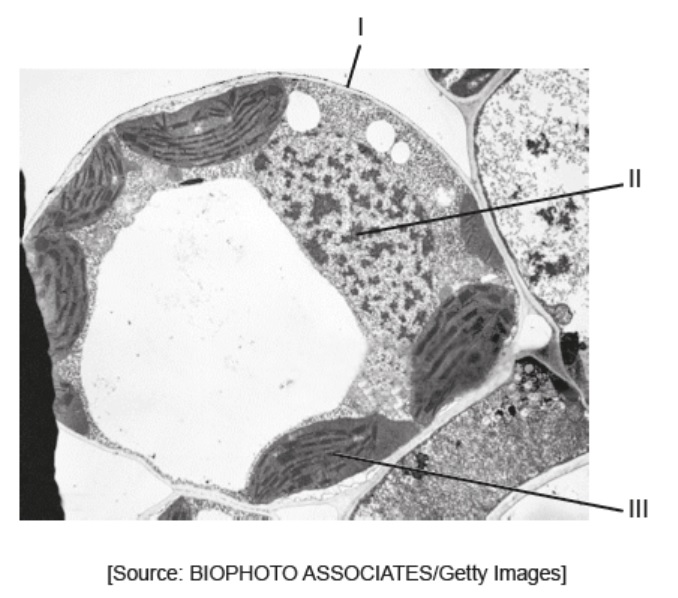
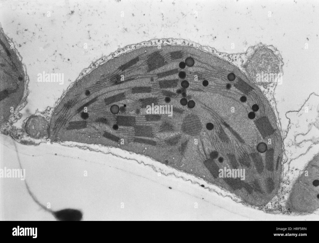
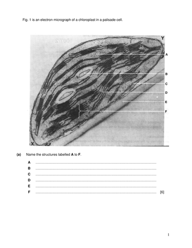
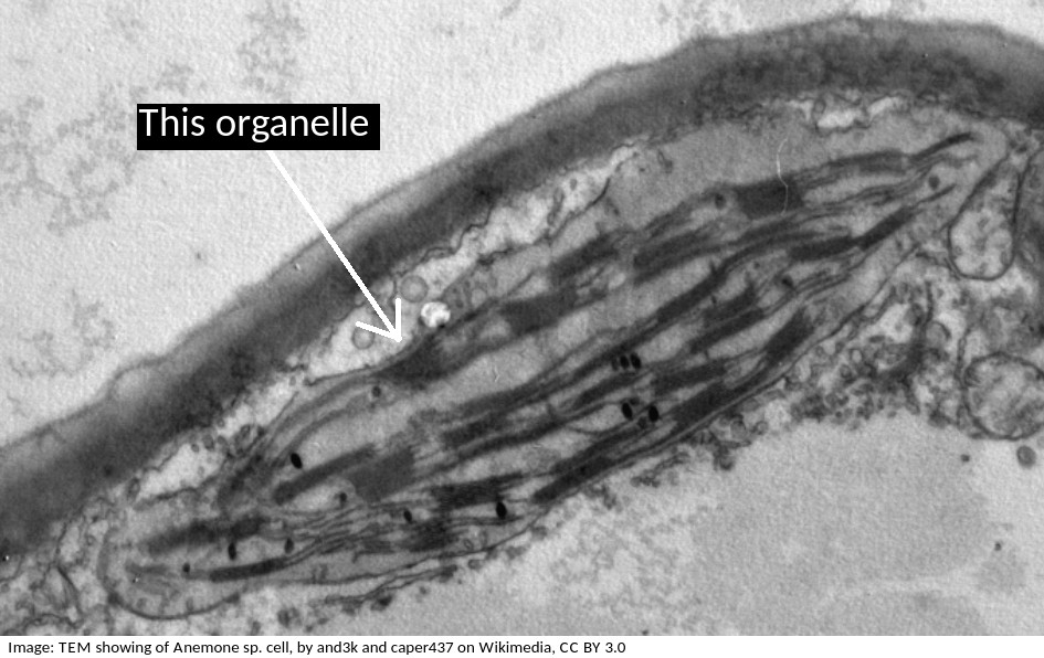
40 labelled electron micrograph of chloroplast
IB Questionbank - examsnap.io 16M.2.HL.TZ0.7a: Draw a labelled diagram of a eukaryotic plant cell as seen in an electron micrograph. 16N.2.SL.TZ0.2a: The image is an electron micrograph. Determine, with a reason, whether the image is of a... 16N.3.SL.TZ0.3a: State from which organ the section was taken. The diagram below represents a section through a chloroplast as seen ... The diagram below represents a section through a chloroplast as seen under the electron microscope. (a) Name the structure labelled D. (b) In which labelled structure do we find chlorophyll molecule. (c) Name the structure labelled, where carbon IV oxide fixation occurs.
Topic 8 2 Photosynthesis Electron micrograph of chloroplast Electron micrograph of chloroplast n n n Book page 170 _____ outer membrane Internal membranes called ____ which is the location of the _____ of the thylakoid called _____ surrounding the thylakoids and inside the double membrane of the chloroplast. This is the location of the _____ that includes the _____ The stroma often contains _____ and ...

Labelled electron micrograph of chloroplast
Chloroplasts - Definition, Structure, Function and Microscopy To view chloroplasts under the microscope, students can use toluidine blue stain to prepare a wet mount. This simply involves the following simple steps: Place a plant sample onto drop of water on a clean glass slide Using a dropper, add a drop of the stain (toluidine blue) on the sample and allow to stand for about a minute Electron Micrographs of Cell Organelles | Zoology This is an electron-micrograph of plastid or chloroplast, which is an integral component of all green plant leaves and is characterized by following features (Fig. 15 & 16): (1) They may be spheroidal, ovoid, stellate or collar shaped and differ in size and number in different cells. Draw a labelled diagram of chloroplast as seen under an electron ... Click here👆to get an answer to your question ️ Draw a labelled diagram of chloroplast as seen under an electron microscope. Name the three major photosynthetic pigments. Solve Study Textbooks Guides. ... Draw a labelled diagram of chloroplast as seen under an electron microscope. Name the three major photosynthetic pigments. Hard. Open in App.
Labelled electron micrograph of chloroplast. Electron Micrographs** Electron Micrographs**. Below is a collection of electron micrographs with labelled subcellular structures that you should be able to identify. Also, be sure to observe any electron micrographs which are made available in the laboratory by the instructor. You should concentrate on the similarities in form that permit identification of the ... Draw a labelled diagram of chloroplast as seen under an electron ... Click here👆to get an answer to your question ️ Draw a labelled diagram of chloroplast as seen under an electron microscope. Name the three major photosynthetic pigments. Join / Login >> Class 11 >> Biology >> Photosynthesis in Higher Plants >> Types of Pigments Involved in Photosynthesis Classical transmission electron microscopy (TEM) led to the formulation ... Thin section electron micrograph of a chemically fixed chloroplast in a young tobacco leaf. The chloroplast lies flat against the plasma membrane and the cell wall (CW) and presents a more or less elliptical outline. The stacked grana thylakoids (GT) are interconnected by non-stacked stroma thylakoids (ST). PDF Chloroplasts Structure and Function Factsheet The biconvex shape of the chloroplast is yet another way of increasing surface area to maximise absorption of light energy Sometimes in the exam you will be presented with an electron micrograph of a chloroplast. Usually, the first question simply asks you to label it. Typical Exam Question Label parts A B & C A B C Answer A - stroma;
Chloroplasts - Biology Pages The electron micrograph above on the right (courtesy of Dr. L. K. Shumway) shows the chloroplast from the cell of a corn leaf. The electron micrograph on the left (courtesy of Kenneth R. Miller) shows the inner surface of a thylakoid membrane. Each particle may represent one photosystem II complex. Plant Cell Nucleus Electron Micrograph : Cell And Organelles Dr Jastrow ... The nucleus (plural = nuclei) figure 7.14 at left a transmission electron micrograph and at right a labeled diagram of a. An electron micrograph of a cell nucleus showing a densely staining nucleolus. Plant cell, electron micrograph 13 plant cells and tissues 29, 30 fiber 11. Plant cells also contain chloroplasts, often have a large, permanent ... Scanning electron microscopy of chloroplast ultrastructure Abstract. A range of fracturing and sectioning techniques are now available which permit intracellular structures to be observed in the scanning electron microscope. One such technique, based on the method of Tanaka (1981), has been used to study chloroplast ultrastructure in Japan laurel, Aucuba japonica. Small pieces of leaves were fixed ... Labeling the Cell Flashcards | Quizlet Label the structures of the plasma membrane and cytoskeleton. Label the membranous organelles. ... Label the transmission electron micrograph of the mitochondrion. Label the transmission electron micrograph of the nucleus. membrane bound organelles. golgi apparatus, mitochondrion, lysosome, peroxisome, rough endoplasmic reticulum ...
Solved Examine this electron micrograph of a chlorplast. - Chegg Question: Examine this electron micrograph of a chlorplast. A. Identify the stack of membranes labeled A. B. Identify the region labeled B. C. Would the production of organic compounds during the light-independent reactions occur in This problem has been solved! See the answer Examine this electron micrograph of a chlorplast. Chloroplast Micrograph Stock Photos and Images - Alamy Transmission electron micrograph of a chloroplast ID: CXWTYX (RF) High magnification micrograph of a chloroplast ID: CXWTYJ (RF) Tomato Chloroplast, TEM ID: HRJNWB (RM) Plant tissue, light micrograph. ID: 2DAMBR2 (RF) Electron microscopy of a vegetal cell showing chloroplast, nucleus, nucleole and cell walls. ID: 2HD77AN (RM) Cell Biology, Chloroplast - University of Florida Electron micrograph of a chloroplast The inner membrane system of the chloroplast is called the thylakoid membranes and the matrix surrounding the thylakoids is called the stroma. Stacks of thylakoids are termed the grana, while the membranes connecting the grana are called the stroma thylakoids. Label This Transmission Electron Micrograph : TEM of chloroplast from ... Provide the labels for the electron micrograph in figure 12.8. Label the transmission electron micrograph of the nucleus. Label the transmission electron micrograph of the nucleus. Transmission electron microscopy (tem) is a microscopy technique in which a beam of electrons is transmitted through a specimen to form an image.
chloroplast and mitochondrion.pdf - D SBA#: Title: Electron micrograph ... sba# electron micrograph of a chloroplast skilled assessed: d (drawing) criteria total marks marks obtained no evidence of shading 1 drawing is a good representation of specimen 1parts of drawings are proportional to parts of specimen (-1 for deviation) 2 drawing is large enough to observe necessary details (occupies at least 50% of page) 1 …
plant cell label electron micrograph Diagram | Quizlet Start studying plant cell label electron micrograph. Learn vocabulary, terms, and more with flashcards, games, and other study tools.
Leaf chloroplast - Radboud Universiteit High-resolution SEM image of the sponge paremchyma cells and the chloroplast © with labels or without labels (172 KB). Printable version of this page in Word or pdf format. TEM view of a single chloroplast and details Chloroplast of tobacco: 1 = cell wall 2 = cytoplasm 3 = vacuole 4 = chloroplast envelope (2 membranes) 5 = tonoplast
Chloroplast- Diagram, Structure and Function Of Chloroplast It is oval or biconvex, found within the mesophyll of the plant cell. The size of the chloroplast usually varies between 4-6 µm in diameter and 1-3 µm in thickness. They are double-membrane organelle with the presence of outer, inner and intermembrane space. There are two distinct regions present inside a chloroplast known as the grana and stroma.
Electron micrograph of isolated chloroplasts with the major organellar ... Electron micrograph of isolated chloroplasts with the major organellar subcompartments labelled. Arrows indicate immunogold-labelled preprotein that is trapped at an intermediate stage of...
Transmission electron microscopic images of chloroplasts and ... For easy organelle identification, a chloroplast (P) and a mitochondrion (M) are labeled. (C-E) Ultrastructure of chloroplasts and mitochondria in cells of the strong PRORP1 RNAi mutant line...
PDF Identifying Organelles from an Electron Micrograph The photograph shown below details chloroplast structure as viewed with a transmission electron microscope Courtesy of Dr. Julian Thorpe - EM & FACS Lab, Biological Sciences University Of Sussex A single Granum Chloroplast envelope visible as two membranes Stroma containing numerous small ribosomes Lamellae connecting different grana
Electron micrographs chloroplast - Big Chemical Encyclopedia (a) Electron micrograph of the thylakoid membranes of maize chloroplasts kindly provided by Dr. D.J. Goodchild, and (b) a diagrammatic representation of the appressed membranes and the non-appressed regions which are directly exposed to the stromal phase of the chloroplast. [Pg.282] Figure 19.1.
6 examine the electron micrograph of a chloroplast a - Course Hero examine the electron micrograph of a chloroplast. (a) identify the stack of membranes labeled a. - stack of thylakoids is granum (b) identify the region labeled b. - inner aqueous fluid, stroma (c) would the production of organic compounds during the light independent reactions occur in region b or on the membranes labeled a. - light …
Refer to the given diagrammatic representation of an electron ... Correct Answer - B Light reactions (or photochemical phase) of photosynthesis mainly occur on the grana thylakoids. Dark reactions (or biosynthetic phase) which involves synthesis of chrbophydrates by C O2 C O 2 fixation, occur in the stroma (or matrix) of chloroplasts.
Draw a labelled diagram of chloroplast as seen under an electron ... Click here👆to get an answer to your question ️ Draw a labelled diagram of chloroplast as seen under an electron microscope. Name the three major photosynthetic pigments. Solve Study Textbooks Guides. ... Draw a labelled diagram of chloroplast as seen under an electron microscope. Name the three major photosynthetic pigments. Hard. Open in App.
Electron Micrographs of Cell Organelles | Zoology This is an electron-micrograph of plastid or chloroplast, which is an integral component of all green plant leaves and is characterized by following features (Fig. 15 & 16): (1) They may be spheroidal, ovoid, stellate or collar shaped and differ in size and number in different cells.
Chloroplasts - Definition, Structure, Function and Microscopy To view chloroplasts under the microscope, students can use toluidine blue stain to prepare a wet mount. This simply involves the following simple steps: Place a plant sample onto drop of water on a clean glass slide Using a dropper, add a drop of the stain (toluidine blue) on the sample and allow to stand for about a minute







![PDF] The Chloroplast Outer Envelope Membrane: The Edge of ...](https://d3i71xaburhd42.cloudfront.net/2d5ca3766698faba516a4a25f0d399ead7e01d62/3-Figure1-1.png)


Post a Comment for "40 labelled electron micrograph of chloroplast"