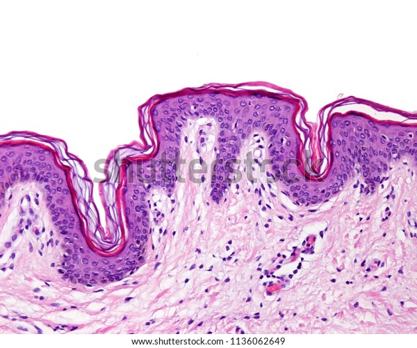
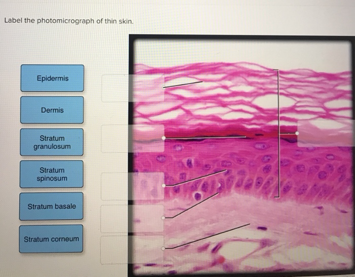
39 label the photomicrograph of thin skin.
Question : Question 31 points Label the photomicrograph of thin skin ... Question 31. A first grade teacher wishes to "shape" her student's writing of the alphabet. The teacher should: a, reward the child whenever the c... Question 31. A neurotransmitter that allows sodium ions to leak into a postsynaptic neuron causes: A) inhibitory postsynaptic damage to the myelin sheath C) excitatory postsynaptic... Label The Photomicrograph Of Thick Skin. - Blogger 1 answer to label the photomicrograph of thin skin. The epidermis, made of closely packed epithelial cells, and the dermis, made of dense, irregular connective tissue . Epidermis Of Thick Skin from eugraph.com The skin is composed of two main layers: Thick skin showing epithelial detail. Practice labeling the layers of the skin.
photomicrographs of thin skin Flashcards | Quizlet photomicrographs of thin skin. STUDY. Flashcards. Learn. Write. Spell. Test. PLAY. Match. Gravity. Created by. Madison_Tacquard. Terms in this set (4) stratum corneum. sebaceous gland. hair follicle. dense irregular CT of the reticular layer of the dermis. Sets found in the same folder. hair structure. 8 terms. Madison_Tacquard.
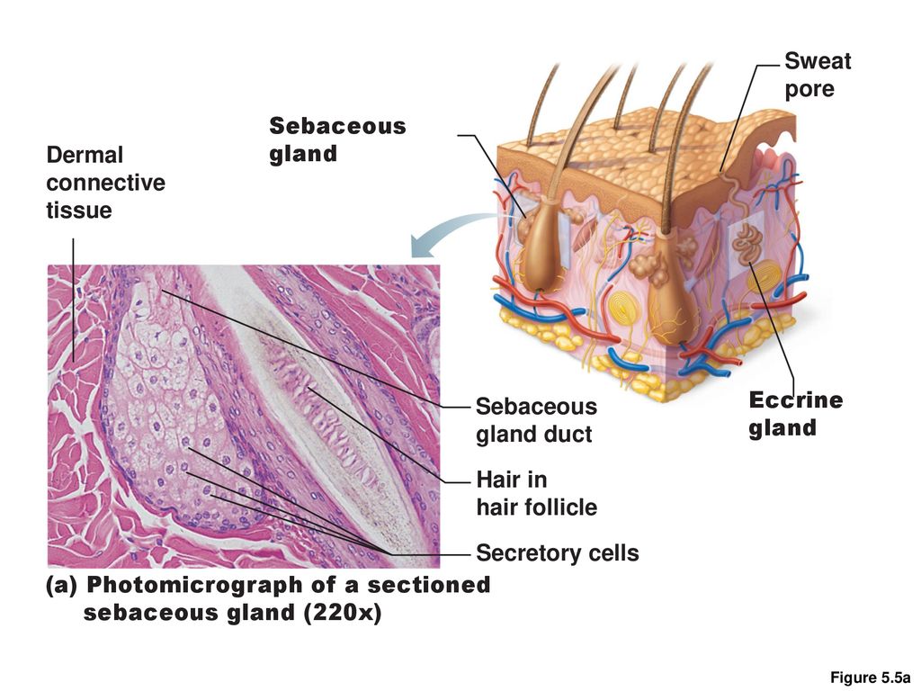
Label the photomicrograph of thin skin.
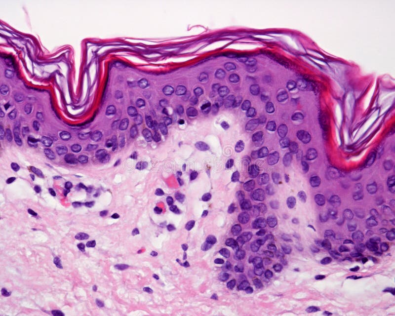
Cambridge International AS & A Level Biology Coursebook ... showing the structure of a generalised plant cell, both as seen with a light microscope. (A generalised cell shows all the structures that may commonly be found in a cell.) Figures 1.6 and 1.7 are photomicrographs. A photomicrograph is a photograph of a specimen as seen with a light microscope. Figure 1.6 shows some human cells. (Solved) - Label the photomicrograph of thin skin. O ... - Transtutors 1 Answer to Label the photomicrograph ... The differences between thick and thin skin - University of Leeds Dermis: Thick skin has a thinner dermis than thin skin, and does not contain hairs, sebaceous glands, or apocrine sweat glands. Thick skin is only found in areas where there is a lot of abrasion - fingertips, palms and the soles of your feet. show labels. This is a picture of an H&E stained section of the epidermis of thin skin.
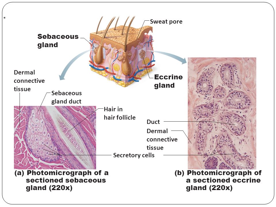
Label the photomicrograph of thin skin.. A&P 1 Exercise_7 Activity 1 & 2 & RYK and UYK.docx - LAB... Apocrine sweat Gland Label the photomicrograph in Figure 7.4. 1. Sebaceous glands 2. Hair follicle 3. Hair root 4. Hair bulb 5. Papilla of hair ... Translucent layer found in thick skin, absent in thin skin. Stratum Spinosum 6. Appears to have thorn-like projections in prepared slides. Reticular Region 7. Label The Photomicrograph Of Thick Skin - Faktor yang Label the photomicrograph of thick skin. 1 answer to label the photomicrograph of thin skin. The epidermis of thick skin has five layers: Hypodermis label the layers of the epidermis in thick skin in figure 7.2. A few layers of cells that are . Apocrine sweat gland label the photomicrograph in figure 7.4. Label the photomicrograph of thick skin. unit 4 lab.docx - LAB Unit 4 EXERCISE 7: The Integumentary... FIGURE 7.4: Diagram of the skin and accessory structures. • apocrine (AP-oh-krin) sweat gland • arrector pili (PIE-lee) muscle • eccrine (EK-rin) sweat gland • hair bulb • hair follicle • hair root • hair shaft • papilla (puh-PILL-uh) of hair • sebaceous (se-BAY-shus) gland 1. Hair shaft 2. Hair root 3. Sebaceous glands 4. Arrector pili muscle 5. Label The Photomicrograph Of Thick Skin / Solved Label The ... - Blogger Label the photomicrograph of thick skin. A few layers of cells that are . Thick skin · stratum basale (also known as s. Solved Label The Photomicrograph Of Thin Skin Chegg Com from d2vlcm61l7u1fs.cloudfront.net. A few layers of cells that are . The stratum lucidum (only found in thick skin), and the stratum corneum.
Photomicrograph of Thin Skin Quiz - PurposeGames.com This is an online quiz called Photomicrograph of Thin Skin. There is a printable worksheet available for download here so you can take the quiz with pen and paper. Your Skills & Rank. Total Points. 0. Get started! Today's Rank--0. Today 's Points. One of us! Game Points. 5. Final Exam A&P 1 Flashcards | Quizlet Label the photomicrograph of thin skin Hair shaft, epidermis, dermal root sheath, sebaceous gland, dermis, hair matrix label the structures of the hair follicle Identify the layers of the epidermis with relation to their location and role in keratinization ... the receptors responsible for olfaction are located in the olfactory epithelium CH 5 Integument - CHAPTER 5 INTEGUMENT Skin (Integument)... - Course Hero Figure 5.2a The main structural features of the skin epidermis. Dermis Stratum spinosum Several layers of keratinocytes unified by desmosomes. Cells contain thick bundles of intermediate filaments made of pre-keratin. Stratum basale Deepest epidermal layer; one row of actively mitotic stem cells; some newly formed cells become part of the more superficial layers. Cambridge International AS & A Level Biology Coursebook … A photomicrograph is a photograph of a specimen as seen with a light microscope. Figure 1.6 shows some human cells. Figure 1.7 shows a plant cell taken from a leaf. Both figures show cells magnified 400 times, which is equivalent to using the high-power objective lens on a light microscope. See also Figures 1.8a and 1.8b for labelled drawings of these figures.
Question : Label the photomicrograph of thin skin. Dermis Duct of ... Expert Answer. 100% (35 ratings) A …. View the full answer. Transcribed image text: Label the photomicrograph of thin skin. Dermis Duct of sebaceous gland Hair Follicle Sebaceous gland Hair Epidermis. Previous question Next question. (Get Answer) - Label The Photomicrograph Of Thick Skin. Stratum ... A) stratum corneum, stratum spinosum, stratum lucidum, stratum granulosum, stratum basale B) stratum basale,... Posted 4 months ago. Q: Procedure Microscopy of Thick Skin obtain a prepared slide of thick skin (which may be labeled "Palmar Skin"), and examine it with the naked eye to get oriented. Once you are oriented, place the slide on the ... Label The Photomicrograph - Mr. Hill's Biology Blog: Our cells "inner skin" Label the photomicrograph of thick skin. Monocyte, erythrocyte, lymphocyte, neutrophil, basophil, eosinophil. (b) for this portion of the problem, we are asked to determine how much total ferrite and cementite form. Photomicrograph is equal to its volume fraction; Label the photomicrograph of thin skin. Layers of the Skin | Anatomy and Physiology I - Lumen Learning Skin that has four layers of cells is referred to as "thin skin.". From deep to superficial, these layers are the stratum basale, stratum spinosum, stratum granulosum, and stratum corneum. Most of the skin can be classified as thin skin. "Thick skin" is found only on the palms of the hands and the soles of the feet.
Anatomy, Skin (Integument), Epidermis - StatPearls - NCBI Bookshelf Skin is the largest organ in the body and covers the body's entire external surface. It is made up of three layers, the epidermis, dermis, and the hypodermis, all three of which vary significantly in their anatomy and function. The skin's structure is made up of an intricate network which serves as the body's initial barrier against pathogens, UV light, and chemicals, and mechanical injury ...
Figure 7.1: Photomicrograph of Skin Diagram | Quizlet Start studying Figure 7.1: Photomicrograph of Skin. Learn vocabulary, terms, and more with flashcards, games, and other study tools.
DiFiore's Atlas of Histology with Functional Correlations ... Enter the email address you signed up with and we'll email you a reset link.
PDF The Integumentary System - Holly H. Nash-Rule, PhD Label the skin structures and areas indicated in the accompanying diagram of thin skin. Then, complete the statements that follow. a. Lamellar granules contain glycolipids that prevent water loss from the skin. b. Fibers in the dermis are produced by fibroblasts .
Theme Pc Anime - Anime Windows 10 11 Themes Page 2 … Share anime theme for windows 7 and windows 8 and anime aimp3 skin. Check out anime based windows 10 desktop themes and choose your favorite anime for your windows theme looks. When it comes to escaping the real worl. Gaming isn't just for specialized consoles and systems anymore now that you can play your favorite video games on your laptop or tablet. However, …
Anatomy and Physiology Homework Chapter 6 Flashcards | Quizlet Label the photomicrograph of thin skin.-Epidermis-Stratum spinosum-Stratum basale-Stratum granulosum-Stratum corneum-Dermis
DiFiore's Atlas of Histology with Functional Correlations (11th … Enter the email address you signed up with and we'll email you a reset link.
Junqueira's Basic Histology Text and Atlas, 14th Edition Enter the email address you signed up with and we'll email you a reset link.
photomicrograph of thick skin Diagram | Quizlet Start studying photomicrograph of thick skin. Learn vocabulary, terms, and more with flashcards, games, and other study tools.
Sebaceous Gland Label The Photomicrograph Of Thin Skin - Integumentary ... The skin and its associated structures, hair, sweat glands and nails make up . 1 answer to label the photomicrograph of thin skin. (b) a photomicrograph of h&e section of thin skin tissue from burnt . Long thin myoepithelial cells are arranged helically around the periphery between the . Dermis duct of sebaceous gland hair follicle sebaceous gland hair epidermis. Label the photomicrograph of thin skin. Part a is a micrograph showing a cross section of thin skin.
Label the photomicrograph in Figure 7.4. Examine a slide of hairy skin ... Label The Photomicrograph Of The Skin And Its Accessory Structures. Sebaceous Gland Duct Of Sebaceous Gland Epidermis Hair Follicle ... Activity 4 Differentiating Sebaceous and Sweat Glands Microscopically Using the slide thin skin with hairs and the photomicrographs of cutaneous glands (Figure 7.6) as a guide, identify sebaceous and eccrine ...
Anime Windows 10 11 Themes Page 2 Themepack Me - Blogger A free desktop customization program for wi. Learn how to make your windows desktop look way more interesting and beautiful with this anime setup. See more ideas about anime, windows themes, online themes. Share anime theme for windows 7 and windows 8 and anime aimp3 skin. Here's 36 best free windows 10 anime theme free download 2022 · 1.
Junqueira's Basic Histology Text and Atlas, 14th Edition Enter the email address you signed up with and we'll email you a reset link.
(Solved) - Label The Photomicrograph Of The Skin And Its Accessory ... FIGURE 7.3 Diagram of the ...
PDF Name the Condition Name the 4 layers of thin skin in both the cartoon and the photomicrograph. Name the 4 layers of thin skin in both the cartoon and the photomicrograph. •Name the Layers of skin and label the dermal papilla and dermis •Name the Layers of skin and label the dermal papilla
The differences between thick and thin skin - University of Leeds Dermis: Thick skin has a thinner dermis than thin skin, and does not contain hairs, sebaceous glands, or apocrine sweat glands. Thick skin is only found in areas where there is a lot of abrasion - fingertips, palms and the soles of your feet. show labels. This is a picture of an H&E stained section of the epidermis of thin skin.
(Solved) - Label the photomicrograph of thin skin. O ... - Transtutors 1 Answer to Label the photomicrograph ...
Cambridge International AS & A Level Biology Coursebook ... showing the structure of a generalised plant cell, both as seen with a light microscope. (A generalised cell shows all the structures that may commonly be found in a cell.) Figures 1.6 and 1.7 are photomicrographs. A photomicrograph is a photograph of a specimen as seen with a light microscope. Figure 1.6 shows some human cells.

















Post a Comment for "39 label the photomicrograph of thin skin."